Ct Scan Pancreatic Cancer Images

The Radiology Assistant Pancreatic Cancer
radiologyassistant.nl
Virtualmedstudent Com Pancreatic Adenocarcinoma
www.virtualmedstudent.com

Malignant Duodenal Lesion Duodenal Tumor Vs Pancreatic Tumor Semiological Radioimaging Characteristics
symbiosisonlinepublishing.com

Pancreatic Cancer Hpblondon Com
www.hpblondon.com

Pancreatic Cancer Ct Wikidoc
www.wikidoc.org

Phase I Study Of Dmot4039a An Antibody Drug Conjugate Targeting Mesothelin In Patients With Unresectable Pancreatic Or Platinum Resistant Ovarian Cancer Molecular Cancer Therapeutics
mct.aacrjournals.org
Using magnetic waves a scanner creates detailed images of the abdomen in particular the area around.

Ct scan pancreatic cancer images. Ct scans help doctors diagnose and treat medical conditions such as pancreatic cancer. Ct or cat stands for computed axial tomography. Computed tomography ct also called computerized axial tomography cat is a procedure used to create 3d body images.
It takes pictures from different angles. Reviewed by benjamin c. When pancreatic cancer is suspected a ct scan is the first imaging study done.
A ct scan is a test that uses x rays and a computer to create detailed pictures of the inside of your body. Pancreatic cancer is one of the most vicious cancers because by the time symptoms start presenting the disease is in an advanced state making prognosis very grim. Using a scope to create ultrasound pictures of your pancreas.
With the ge discovery petct 600 scanner a four dimensional ct scanner that produces detailed cross sectional x ray images of structures within the body our radiologists are better able to plan treatment in accordance with patients. Computed tomography ct scan. The ct scan makes detailed cross sectional images of your body.
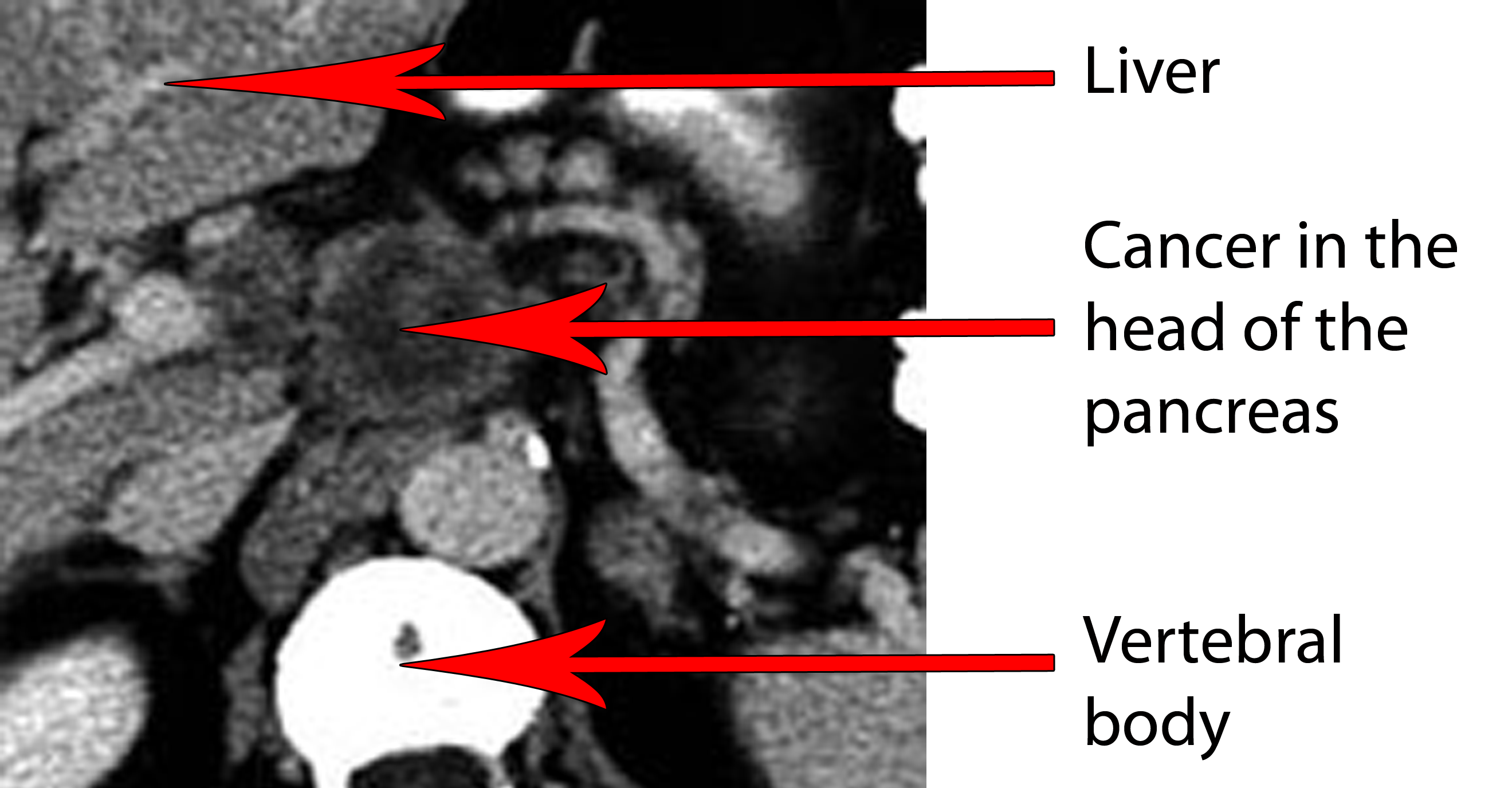
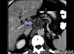
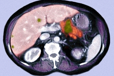
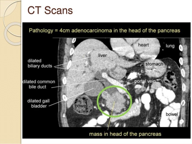
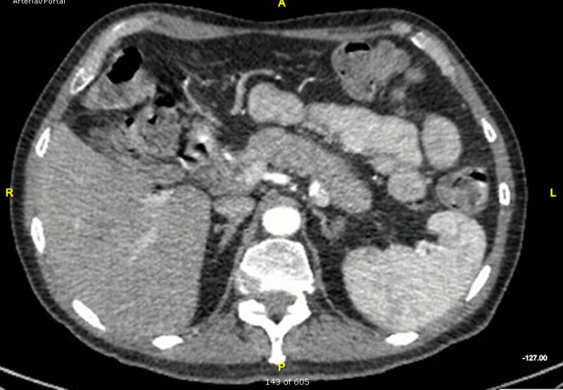
Magnetic resonance imaging mri. Picture of pancreatic cancer an abdominal ct scan shows a small vaguely seen 2 cm pancreatic adenocarcinoma mass causing obstruction of both the common bile duct cbd and pancreatic duct pd. The computer puts them together to make a 3 dimensional 3d image.
These tests help your doctors visualize your internal organs including the pancreas. A ct scan for pancreatic cancer uses x ray images to present detailed images of the pancreas. Before a ct scan doctors give patients a contrast dye as a drink or iv.
You usually have a ct scan in the x ray radiology department as an. Wedro md faaem on july 28 2008. Ct scans are often used to diagnose pancreatic cancer because they can show the pancreas fairly clearly.
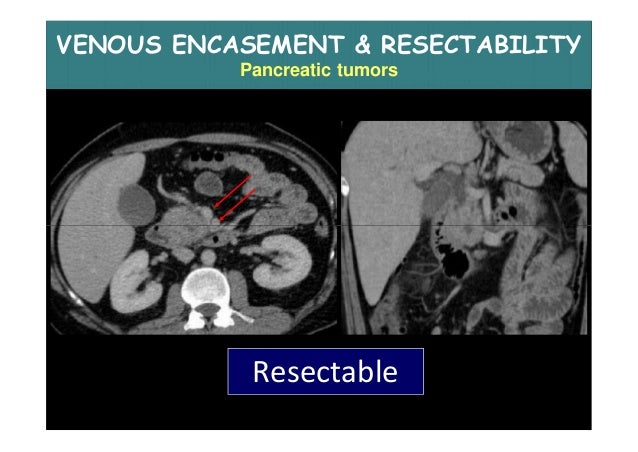
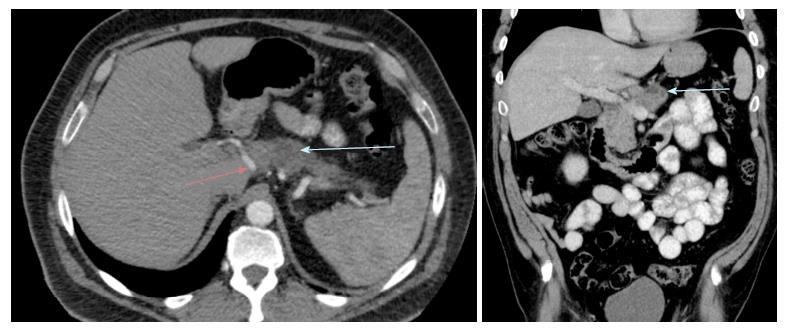
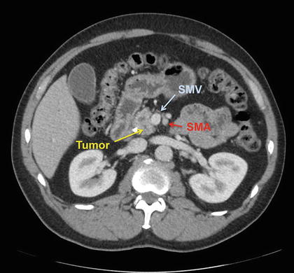
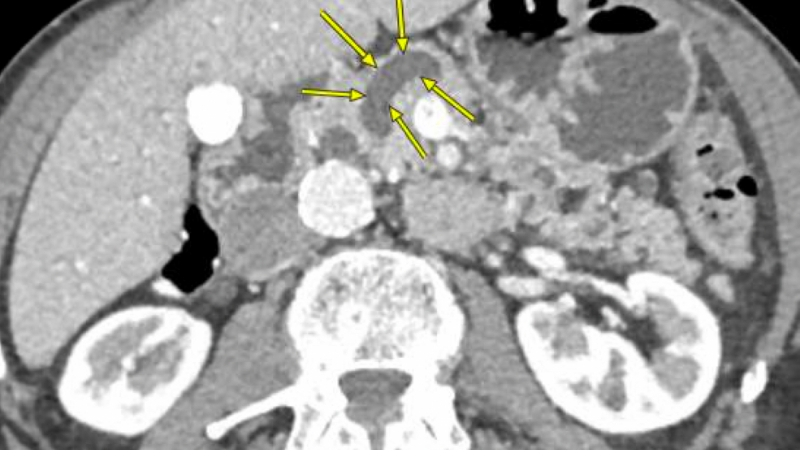
Pancreatic ductal adenocarcinoma makes up the vast majority 90 of all pancreatic neoplasms and remains a disease with a very poor prognosis and high morbidity. These pancreatic head tumors can be missed even on a technically excellent ct and therefore a negative ct scan in a patient with a strong suspicion for pancreatic head cancer requires additional imaging with endoscopic ultrasound. On imaging it usually presents as a hypodense mass on ct that is poorly marginated which may encase vessels and the common bile duct.
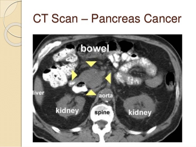
They can also help show if cancer has spread to organs near the pancreas as well as to lymph nodes and distant organs.

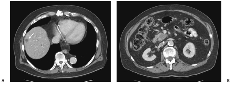
Pancreatic Cancer With Liver And Lung Metastases Pancreas Case Studies Ctisus Ct Scanning
ctisus.com
Hereditary Vs Familial Pancreatic Cancer Associated Genetic Syndromes And Clinical Perspective
www.cancernetwork.com
Use Of A Novel Herbal Medicine In A 75 Year Old Woman With Multi Metastatic Pancreatic Cancer A Case Report And Review Of The Literature
www.spandidos-publications.com

Ct Scans Of Pancreatic Cancer Patient Ct Scan Imaging At Baseline Download Scientific Diagram
www.researchgate.net

Test Shown To Improve Accuracy In Identifying Precancerous Pancreatic Cysts
www.hopkinsmedicine.org
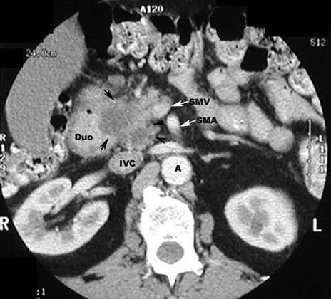
Imaging Preoperatively For Pancreatic Adenocarcinoma Pietryga Journal Of Gastrointestinal Oncology
jgo.amegroups.com

Imaging To Cure Cancer The Contribution Of Radiology Pharmacyte Biotech
pharmacyte.com

Epos Trade
epos.myesr.org
Pancreatic Cancer Ct Scan Medlineplus Medical Encyclopedia Image
medlineplus.gov

46 Year Old Woman With Pancreatic Cancer And Prior Distal Download Scientific Diagram
www.researchgate.net
Imaging Of The Pancreas
www.slideshare.net
Ct Scan Of Patient With Distal Pancreatic Tumor Cancer Surgery Associates
drdonaldmccain.com

Cancers Free Full Text Current State Of Surgical Management Of Pancreatic Cancer Html
www.mdpi.com

Pancreatic Cancer Suspected And Staging
imagingpathways.health.wa.gov.au

Pancreatic Cancer With Liver And Bone Metastases Pancreas Case Studies Ctisus Ct Scanning
www.ctisus.com

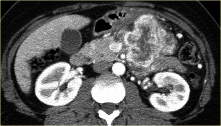
Ct Scan Of The Abdomen Showing The Pancreatic Tumor And The Left Renal Download Scientific Diagram
www.researchgate.net

Pancreatic Cancer Ct Scan Stock Image C029 4650 Science Photo Library
www.sciencephoto.com

Ct Imaging Of Pancreatic Cancer Accessmedicine Network
www.accessmedicinenetwork.com
Pancreas Cancer
www.slideshare.net

Pancreatic Cancer Management Demands Teamwork Multidisciplinary Approach
www.auntminnieeurope.com
Cureus Solid Pseudopapillary Tumor Of The Pancreas An Unusual Cause Of Abdominal Pain
www.cureus.com
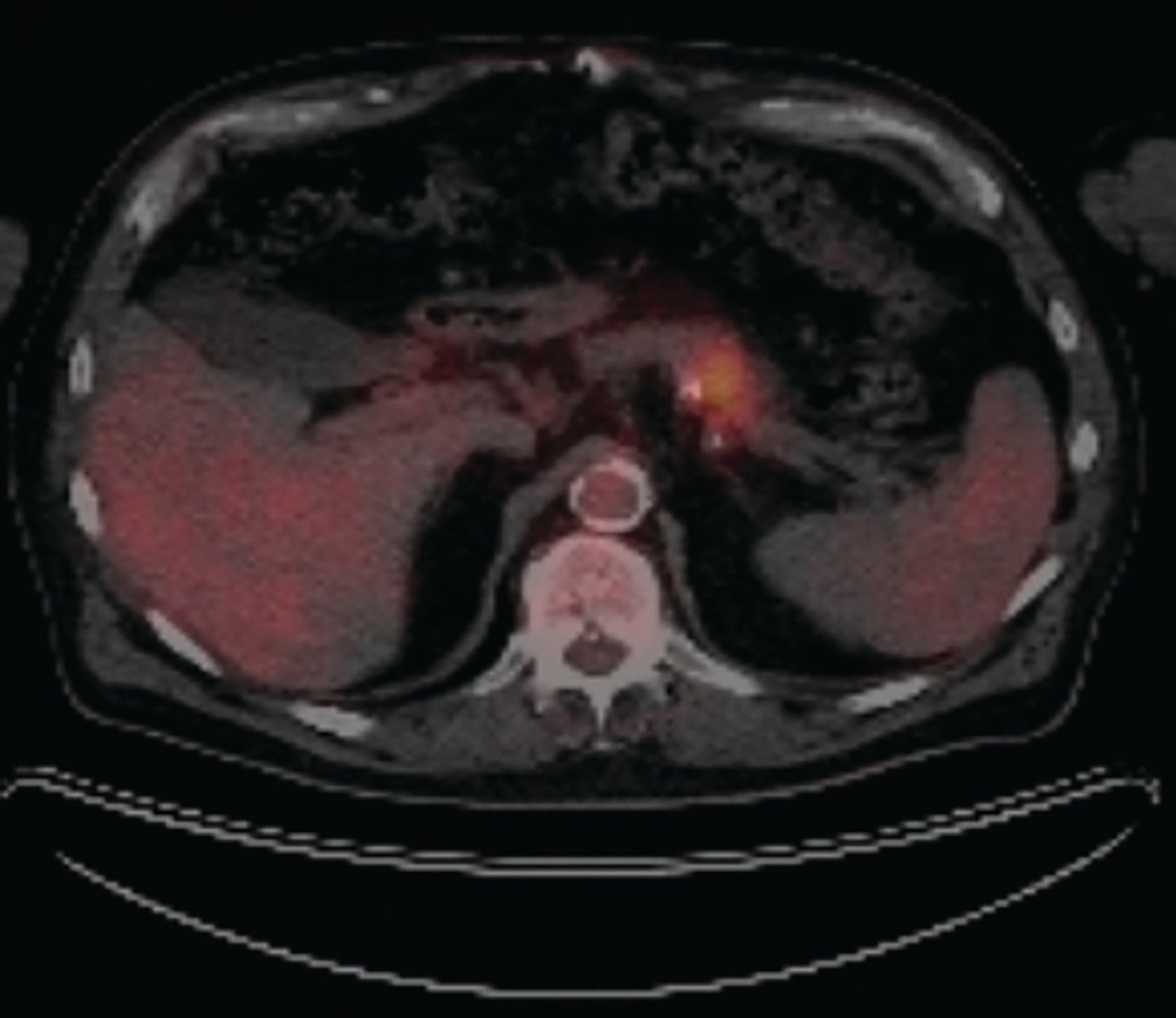
Cross Modality Pet Ct And Contrast Enhanced Ct Imaging For Pancreatic Cancer
www.wjgnet.com
Resection Of Pancreatic Ductal Adenocarcinoma With Synchronous Distant Metastasis Is It Worthwhile World Journal Of Surgical Oncology Full Text
wjso.biomedcentral.com
Immuno Pet Shows Promise For Detecting And Treating Pancreatic Tumors
medicalxpress.com
Anatomic Definitions Of Borderline Resectable Pancreatic Cancer Abdominal Key
abdominalkey.com
Choriocarcinoma Involving The Pancreas As First Manifestation Of A Metastatic Regressing Mixed Testicular Germ Cell Tumor Modern Pathology
www.nature.com

Cystic Tumours Hpblondon Com
www.hpblondon.com
Pancreatic Cancer Patients Should Be Offered Early Scans To Avoid Unnecessary Surgery Says Nice News And Features News Nice
www.nice.org.uk

Hepatic Metastases From Pancreatic Cancer Radiology Case Radiopaedia Org
radiopaedia.org

Pancreatic Cancer Diagnosis And Management American Family Physician
www.aafp.org
1
encrypted-tbn0.gstatic.com

Pancreatic Cancer Diagnosis And Management American Family Physician
www.aafp.org
Multimodality Treatment By Folfox Plus Hifu In A Case Of Advanced Pancreatic Carcinoma A Case Report Insight Medical Publishing
pancreas.imedpub.com
Challenges In Diagnosis Of Pancreatic Cancer
www.wjgnet.com
Https Encrypted Tbn0 Gstatic Com Images Q Tbn 3aand9gctq9hjsspvlyt1crjnqi4j Ul91vpqrjz6gnp2wlupzd7nv7kxe Usqp Cau
encrypted-tbn0.gstatic.com
Clinical Review Management Of Pancreatic Cancer Gponline
www.gponline.com

Umbilical Mass As The Sole Presenting Symptom Of Pancreatic Cancer A Case Report
www.scielo.br
Pancreatic Cancer With Liver Metastases Pancreas Case Studies Ctisus Ct Scanning
ctisus.com
Imaging For Pancreatic Ductal Adenocarcinoma Horvat Chinese Clinical Oncology
cco.amegroups.com

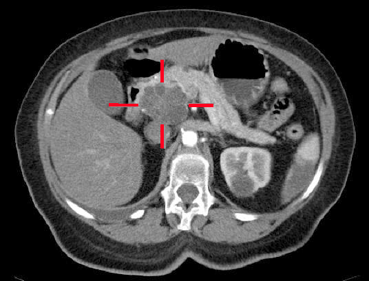
Staging Ct Scan Tumor Of The Tail Of The Pancreas Invading The Spleen Download Scientific Diagram
www.researchgate.net
Pancreatic Cancer With Double Duct Sign Pancreas Case Studies Ctisus Ct Scanning
www.ctisus.com

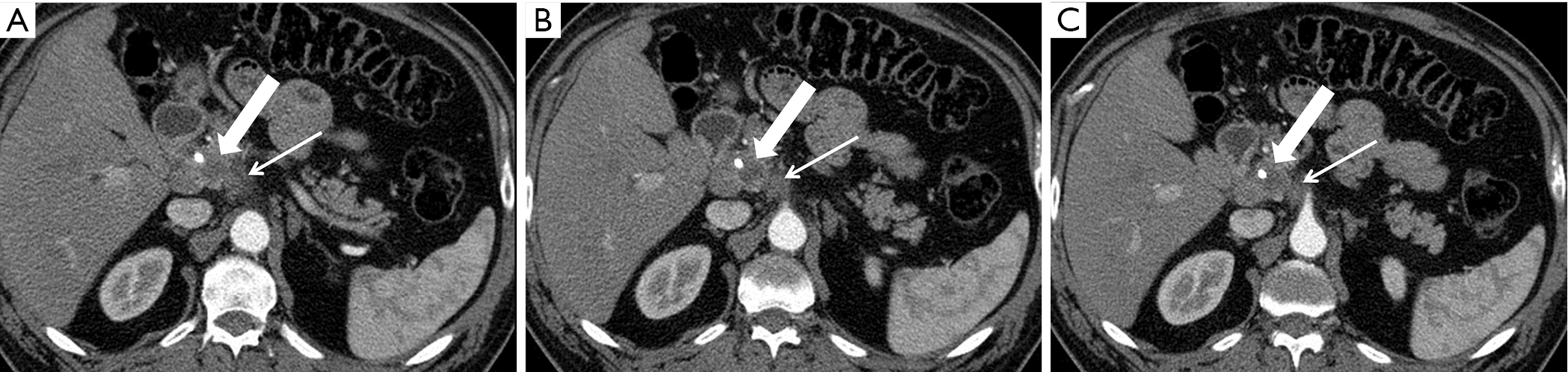
Endoscopic Ultrasound For Detecting Small Pancreatic Tumor Missed By Computed Tomography Sciencedirect
www.sciencedirect.com

About Pancreatic Cancer Lustgarten Foundation
lustgarten.org

Radiologist Apologises For Missing Pancreatic Cancer In Ct Scan New Zealand Doctor
www.nzdoctor.co.nz

Anatomic Definitions Of Borderline Resectable Pancreatic Cancer Abdominal Key
abdominalkey.com

Pancreas Ductal Adenocarcinoma And Its Mimics Review Of Cross Sectional Imaging Findings For Differential Diagnosis
www.jbsr.be
Assessment Of Liver Lesions
www.medscape.org
Https Encrypted Tbn0 Gstatic Com Images Q Tbn 3aand9gcrqivmkzbshivuexviy9dar1tazqsdywqwkvlhvjcbs Sxuljw8 Usqp Cau
encrypted-tbn0.gstatic.com
Https Encrypted Tbn0 Gstatic Com Images Q Tbn 3aand9gcru5vextjpq6bmxsviukzia39qjhkop Al4pmg8utbphirngfjr Usqp Cau
encrypted-tbn0.gstatic.com
Imaging Diagnosis And Staging Of Pancreatic Ductal Adenocarcinoma A Comprehensive Review Insights Into Imaging Full Text
insightsimaging.springeropen.com

Contrast Enhanced Ct Scan Demonstrating A Resectable Adenocarcinoma Of Download Scientific Diagram
www.researchgate.net
Progress Slow Against Pancreatic Cancer
medicalxpress.com
Imaging Diagnosis And Staging Of Pancreatic Ductal Adenocarcinoma A Comprehensive Review Insights Into Imaging Full Text
insightsimaging.springeropen.com
Challenges In Diagnosis Of Pancreatic Cancer
www.wjgnet.com

Diagnosis Ct Scan
www.pathology.jhu.edu

Umbilical Mass As The Sole Presenting Symptom Of Pancreatic Cancer A Case Report
www.scielo.br
18f Fdg Pet Ct Imaging Of The Pancreas Spectrum Of Diseases Insight Medical Publishing
pancreas.imedpub.com
Staging Investigations For Pancreatic Cancer Diagnosis Treatment Surgery
www.pancreaticcancer.co.uk
Pancreatic Cancer Ct Scan Stock Image C001 8039 Science Photo Library
www.sciencephoto.com

Ct Volume Perfusion Imaging In A Case Of Suspected Pancreatic Cancer Siemens Healthineers Colombia
www.siemens-healthineers.com
Anatomic Definitions Of Borderline Resectable Pancreatic Cancer Abdominal Key
abdominalkey.com

Pancreatic Cancer Slow Progression In The Early Stages Sciencedirect
www.sciencedirect.com
Imaging Preoperatively For Pancreatic Adenocarcinoma Pietryga Journal Of Gastrointestinal Oncology
jgo.amegroups.com

Imaging To Cure Cancer The Contribution Of Radiology Pharmacyte Biotech
pharmacyte.com

Pancreatic Cancer Entry 3 Diagnosing Pancreatic Cancer
nppancreaticcancer.blogspot.com
Computed Axial Tomography Cat Or Ct Scan Pancreatic Cancer Action Network
www.pancan.org
The Optimal Rx For Pancreatic Cancer Stop It Before It Starts
www.cancernetwork.com

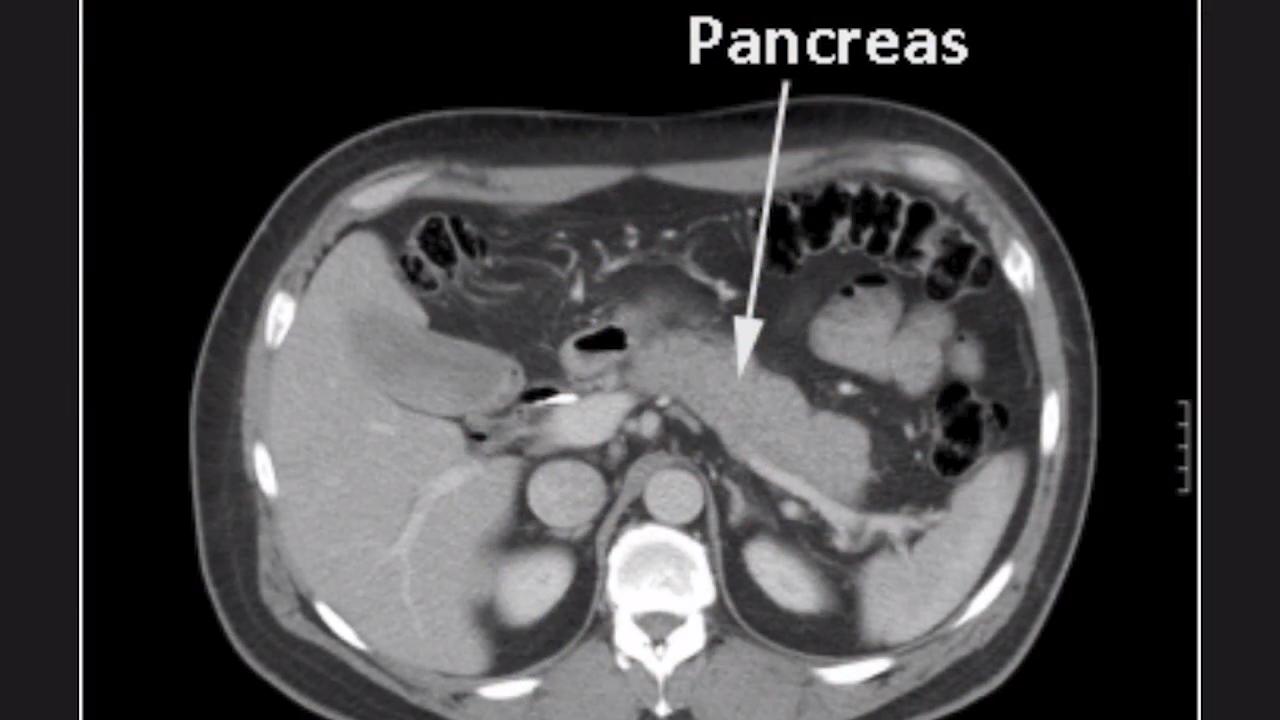
Anatomy Of The Pancreas
www.aboutcancer.com
Video Advances In Pancreatic Ct And Mr Imaging Imaging Technology News
www.itnonline.com
Staging Investigations For Pancreatic Cancer Diagnosis Treatment Surgery
www.pancreaticcancer.co.uk
Pancreas Cancer
www.slideshare.net
9 Liver Metastasis From Primary Pancreatic Cancer Radiology Key
radiologykey.com

Pancreas Cancer Ct Scan Stock Image M134 0463 Science Photo Library
www.sciencephoto.com

Figure 2 From Borderline Resectable Pancreatic Cancer Semantic Scholar
www.semanticscholar.org

5 Ct Scans Of Patients With Borderline Resectable Pancreatic Cancer Download Scientific Diagram
www.researchgate.net
Imaging Preoperatively For Pancreatic Adenocarcinoma Pietryga Journal Of Gastrointestinal Oncology
jgo.amegroups.com

Accessmedicine S Image Of The Week Ct Findings Associated With Pancreatic Cancer Accessmedicine Network
www.accessmedicinenetwork.com

Biology And Management Of Pancreatic Cancer Postgraduate Medical Journal
pmj.bmj.com

Pancreatic Cancer Picture Image On Medicinenet Com
www.medicinenet.com
Pancreatic Cancer Oncology Medbullets Step 2 3
step2.medbullets.com

Novel Mechanism For Anti Cancer Drug Immunopaedia
www.immunopaedia.org.za

Pancreatic Head Carcinoma Radiology Case Radiopaedia Org
radiopaedia.org

Malignant Duodenal Lesion Duodenal Tumor Vs Pancreatic Tumor Semiological Radioimaging Characteristics
symbiosisonlinepublishing.com

Diagnosing Pancreatic Cancer
www.independentnurse.co.uk

Pancreatic Cancer Suspected And Staging
imagingpathways.health.wa.gov.au
The Radiology Assistant Pancreatic Cystic Lesions
radiologyassistant.nl

Pancreas Ductal Adenocarcinoma And Its Mimics Review Of Cross Sectional Imaging Findings For Differential Diagnosis
www.jbsr.be
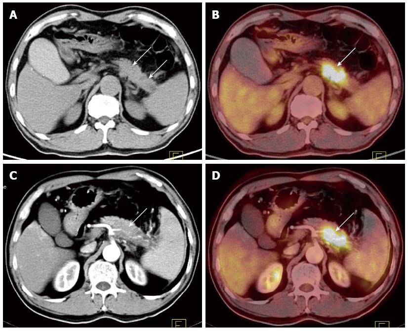
Cross Modality Pet Ct And Contrast Enhanced Ct Imaging For Pancreatic Cancer
www.wjgnet.com

Pitfalls And Pearls In The Ct Diagnosis Of Pancreatic Cancer Youtube
www.youtube.com
Possible Pancreatitis Or Could It Be Cancer Ueg United European Gastroenterology
ueg.eu

Ct Scan For Evaluation Of Abdominal Pain 8 Years After Diagnosis Of Download Scientific Diagram
www.researchgate.net
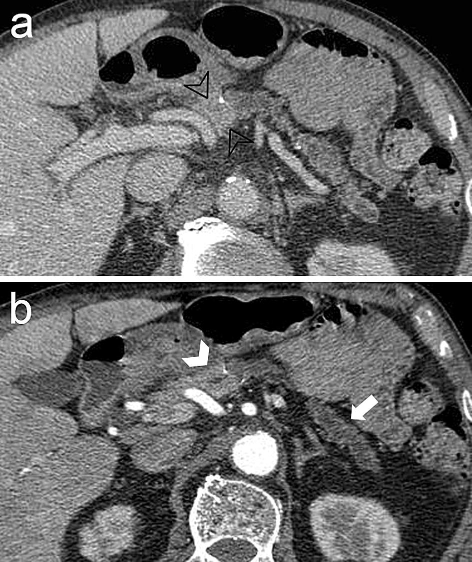
Figure 5 Ct And Mr Features That Can Help To Differentiate Between Focal Chronic Pancreatitis And Pancreatic Cancer Springerlink
link.springer.com

Endoscopic Ultrasound For Detecting Small Pancreatic Tumor Missed By Computed Tomography Sciencedirect
www.sciencedirect.com

Pancreatic Cancer Symptoms Diagnosis And Treatment Cancer Advice
www.canceradvice.co.uk

New Page 2
www.stritch.luc.edu
Earlier More Frequent Removal Of Some Pancreatic Cysts May Decrease Cancer Risk For Some Patients
www.hopkinsmedicine.org

Scientists Prod Immune Cells To Attack Pancreatic Cancer Newsroom
newsroom.uw.edu

Pancreatitis Physiopedia
www.physio-pedia.com
