Lung Cancer Ct Scan Images Database

From Ct Lung Data To Lung Cancer Diagnosis Top Ct Lung Dataset From Download Scientific Diagram
www.researchgate.net

Implementation Of Lung Cancer Screening In Europe Challenges And Potential Solutions Summary Of A Multidisciplinary Roundtable Discussion Esmo Open
esmoopen.bmj.com

Ct Scan Database Of 1000 Sets Was Created For Teaching Ai To Diagnose Covid 19
medicalxpress.com

Radiation Induced Lung Injury Pulmonology Advisor
www.pulmonologyadvisor.com
Ct Imaging Of The 2019 Novel Coronavirus 2019 Ncov Pneumonia Imaging Technology News
www.itnonline.com

Lung Cancer The Bmj
www.bmj.com
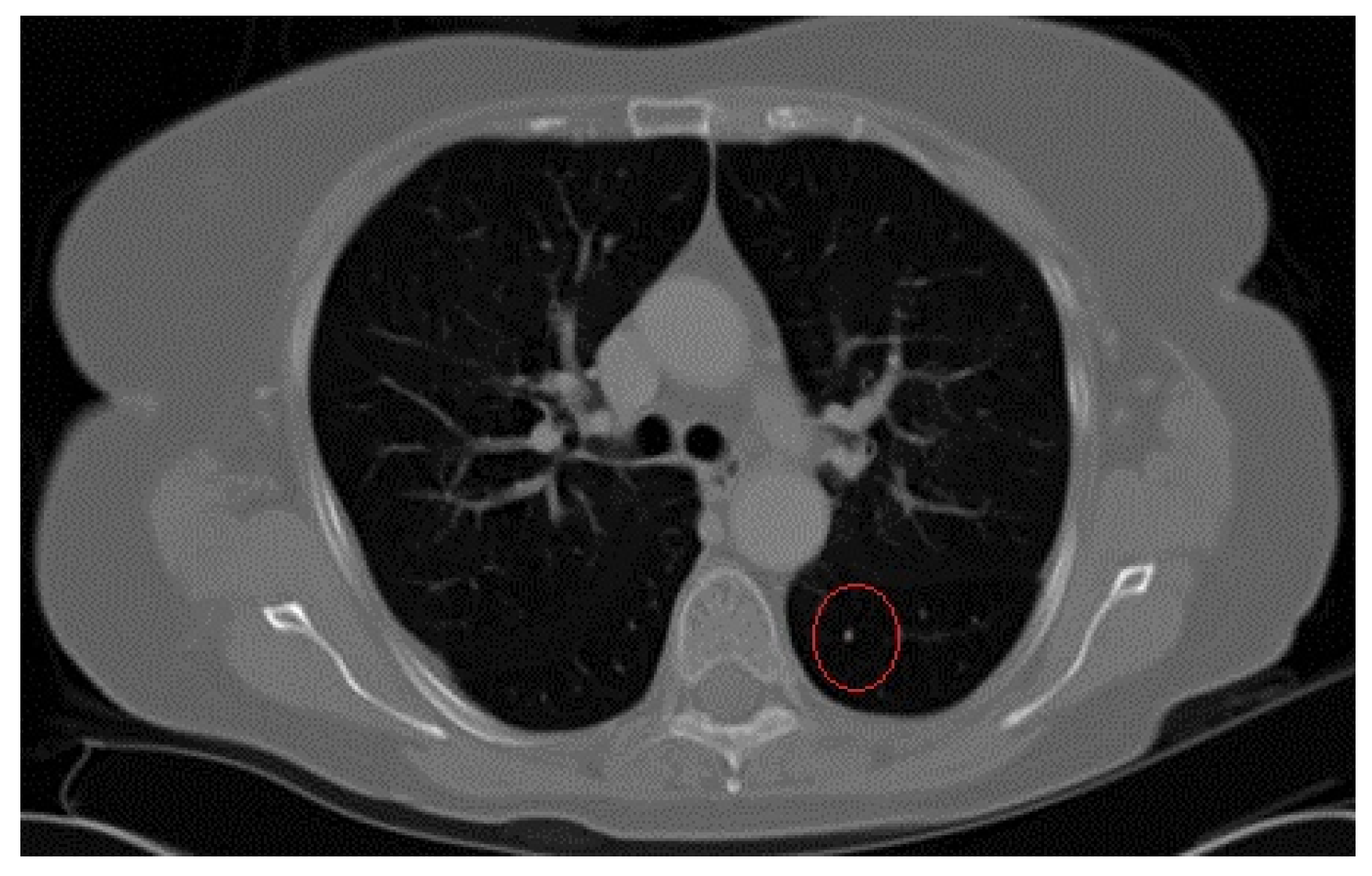
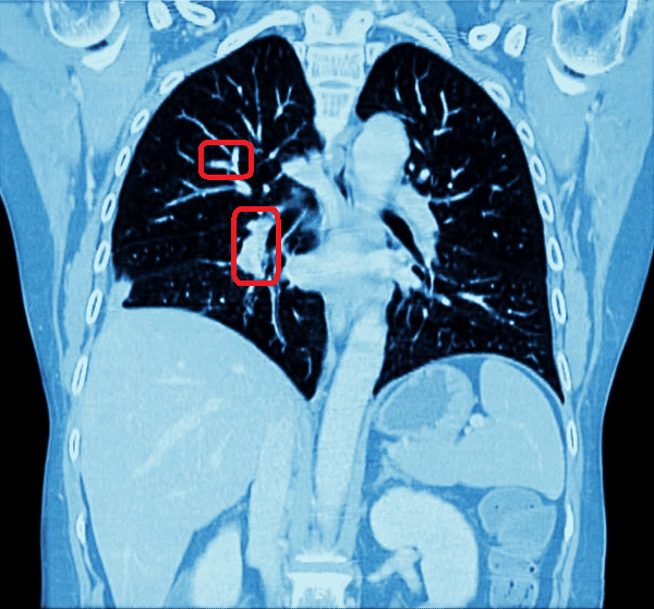
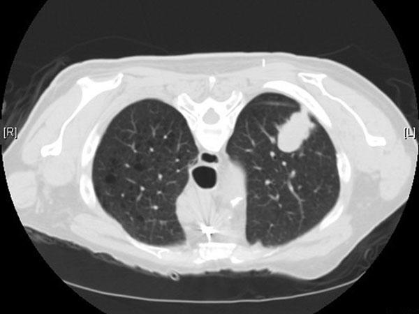
It can give more information about any abnormalities nodules or lesions small abnormal areas in the lungs that were.
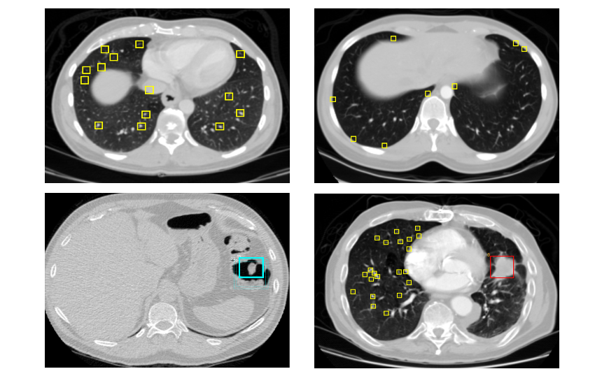
Lung cancer ct scan images database. This database is still under development. The database currently consists of an image set of 50 low dose documented whole lung ct scans for detection. It is a web accessible international resource for development training and evaluation of computer assisted diagnostic cad methods for lung cancer detection and diagnosis.
Lung image database consortium lidc reference image database to evaluate response rider breast mri. The lung image database consortium image collection lidc idri consists of diagnostic and lung cancer screening thoracic computed tomography ct scans with marked up annotated lesions. 50 cases of low dose thin slice chest ct images with annotations for small nodules public database to address drug response.
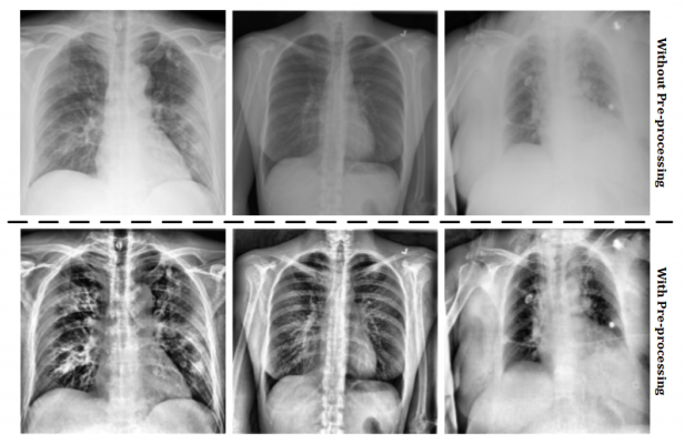
An alternative format for the ct data is dicom dcm. The ct scans were obtained in a single breath hold with a 125 mm slice thickness. A computed tomography scan ct scan also called a cat scan uses computer controlled x rays to create images of the body.
Eclap public database of whole lung ct images. The rider lung ct collection was constructed as part of a study to evaluate the variability of tumor unidimensional bidimensional and volumetric measurements on same day repeat computed tomographic ct scans in patients with nonsmall cell lung cancer. Ct scan database of 1000 sets was created for teaching ai to diagnose covid 19 the largest ct scan database in the world with covid 19 features has been collected in russia.
The locations of nodules detected by the radiologist are also provided. Nih database of 100000 chest x rays. However a radiograph and a ct scan show different types of information.
Over 100 cases of ct chest images illustrating the spectrum of nodule presentations together with a range of computer analysis methods. Although an experienced radiologist can get a sense for the approximate three dimensional location of a tumor from a radiograph in general a plain. Thirty two patients with nonsmall cell lung cancer each of whom underwent two ct scans of the chest within 15 minutes by.
A ct scan takes a cross sectional and a more detailed image of the lung. Images associated clinical data annotations and diagnoses.

Steps In Differential Imaging Our Database That Was Used In This Study Download Scientific Diagram
www.researchgate.net
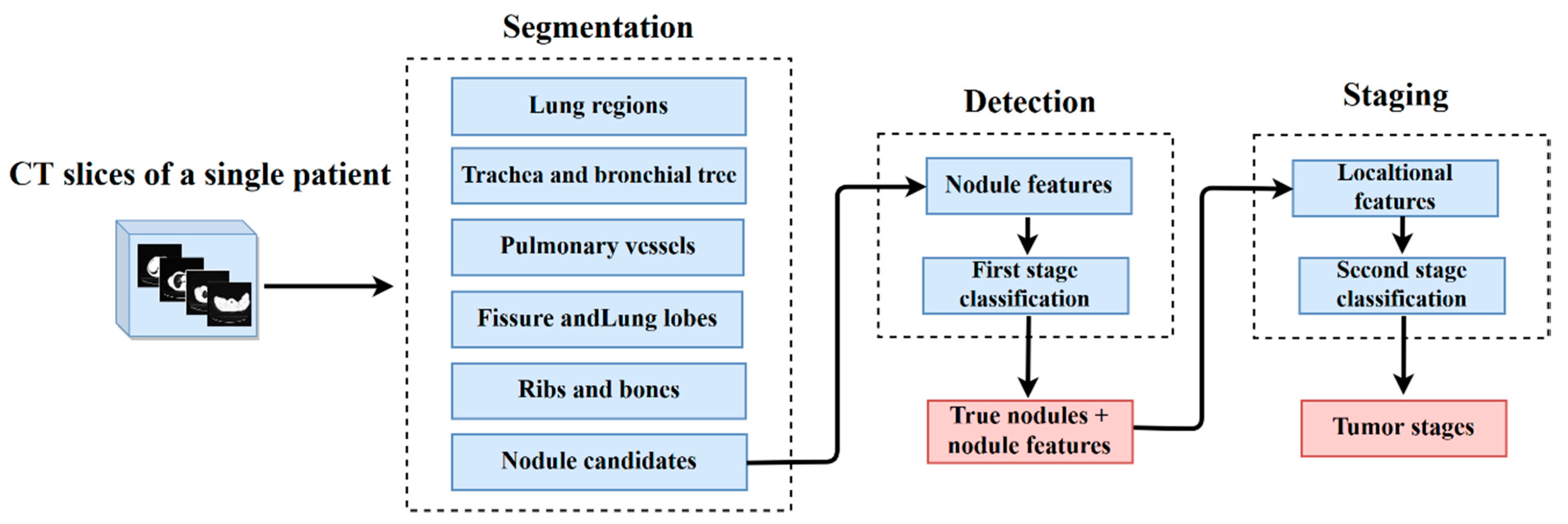
Applied Sciences Free Full Text Automatic Detection And Staging Of Lung Tumors Using Locational Features And Double Staged Classifications Html
www.mdpi.com
Automated System For Lung Nodules Classification Based On Wavelet Feature Descriptor And Support Vector Machine Biomedical Engineering Online Full Text
biomedical-engineering-online.biomedcentral.com

Imaging Techniques In Lung Cancer European Respiratory Society
breathe.ersjournals.com

Pdf Lung Cancer Detection On Ct Scan Images A Review On The Analysis Techniques
www.researchgate.net
Is Lung Cancer Better Detected From An X Ray Or Ct Scan
www.itnonline.com
Using Lung X Rays To Diagnose Covid 19 Imaging Technology News
www.itnonline.com
Applied Sciences Free Full Text Using Double Convolution Neural Network For Lung Cancer Stage Detection Html
www.mdpi.com
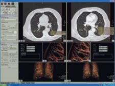
Automated Chest Ct Scan Analysis With Deep Learning Classifier
www.rsipvision.com

Ct Databases Aimed At Lung Imaging Research
www.auntminnie.com

Radiologists Sharing Ct Scans X Rays In Global Effort To Prevent Covid 19 Deaths Abc News
www.abc.net.au

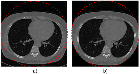
Standard Dose Vs Low Dose Ct Protocols In The Evaluation Of Localized Lung Lesions Capability For Lesion Characterization Ilead Study Sciencedirect
www.sciencedirect.com

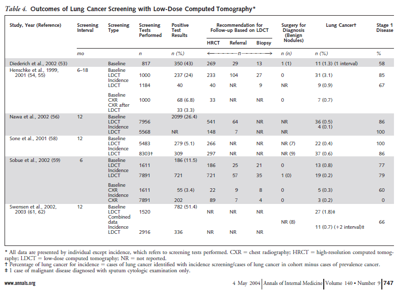
Mass Screening For Lung Cancer With Mobile Spiral Computed Tomography Scanner The Lancet
www.thelancet.com

Uk Lung Cancer Screening Ukls Trial Recruitment And Implementation Download Scientific Diagram
www.researchgate.net

Lung Cancer Screening Wikipedia
en.wikipedia.org

Ct Databases Aimed At Lung Imaging Research
www.auntminnie.com
2nd Place Solution For The 2017 National Datascience Bowl Julian De Wit Freelance Software Machine Learning Engineer
juliandewit.github.io
U S Lung Cancer Screening Experience National Cancer Institute
www.cancer.gov

Limited Stage Small Cell Lung Carcinoma Wikipedia
en.wikipedia.org

Slow Growing Lung Cancer As An Emerging Entity From Screening To Clinical Management European Respiratory Society
erj.ersjournals.com
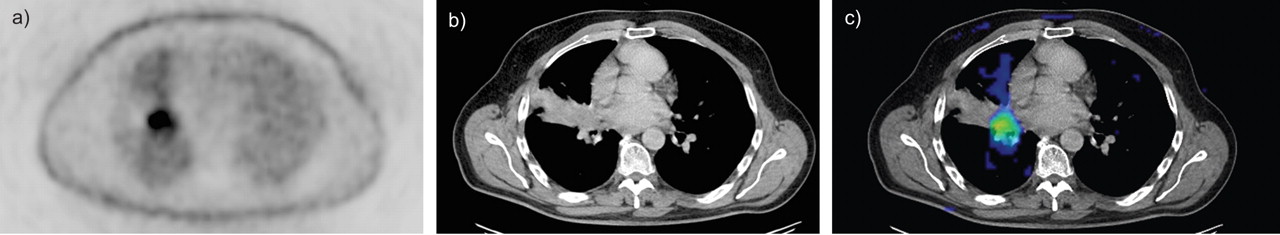
Integrated Pet Ct In The Staging Of Nonsmall Cell Lung Cancer Technical Aspects And Clinical Integration European Respiratory Society
erj.ersjournals.com
Automated System For Lung Nodules Classification Based On Wavelet Feature Descriptor And Support Vector Machine Biomedical Engineering Online Full Text
biomedical-engineering-online.biomedcentral.com
Development And Outcomes Of A Comprehensive Multidisciplinary Incidental Lung Nodule And Lung Cancer Screening Program Bmc Pulmonary Medicine Full Text
bmcpulmmed.biomedcentral.com

Lung Cancer Screening Requires Effective Risk Stratification Pulmonology Advisor
www.pulmonologyadvisor.com

Stages 2 3 And 4 Of Lung Cancer Download Scientific Diagram
www.researchgate.net

Small Cell Lung Cancer Cancer Therapy Advisor
www.cancertherapyadvisor.com
Frontiers Lung Nodule Detection Via Deep Reinforcement Learning Oncology
www.frontiersin.org
Https Www Sciencedirect Com Science Article Pii S1877050917327801 Pdf Md5 B3800b5565e72be67e25327a3e66171d Pid 1 S2 0 S1877050917327801 Main Pdf Valck 1
Small Cell Lung Cancer Sclc
www.slideshare.net
Welcome To The Cancer Imaging Archive The Cancer Imaging Archive Tcia
www.cancerimagingarchive.net

The Imaging Viewpoint How Imaging Affects Determination Of Progression Free Survival Clinical Cancer Research
clincancerres.aacrjournals.org

Clinical Presentation Diagnosis And Management Of Typical And Atypical Bronchopulmonary Carcinoid Mdedge Hematology And Oncology
www.mdedge.com

Ct Medical Images Kaggle
www.kaggle.com
Integrated Pet Ct In The Staging Of Nonsmall Cell Lung Cancer Technical Aspects And Clinical Integration European Respiratory Society
erj.ersjournals.com
Https Encrypted Tbn0 Gstatic Com Images Q Tbn 3aand9gct2cmzyx4oo1z Uzizk0hx8hwtr0gzpnfnd4eyw Mmijgafwcqa Usqp Cau
encrypted-tbn0.gstatic.com

Standard Dose Vs Low Dose Ct Protocols In The Evaluation Of Localized Lung Lesions Capability For Lesion Characterization Ilead Study Sciencedirect
www.sciencedirect.com
Imaging Techniques In Lung Cancer European Respiratory Society
breathe.ersjournals.com

Lung Cancer X Rays Fail To Detect Almost A Quarter Of Cases
www.medicaldevice-network.com
Effective And Reliable Framework For Lung Nodules Detection From Ct Scan Images Scientific Reports
www.nature.com

A Computer Aided Pipeline For Automatic Lung Cancer Classification On Computed Tomography Scans
www.hindawi.com
A Patient With Newly Diagnosed Advanced Egfr Mutated Non Small Cell Lung Cancer
www.cancernetwork.com
Deep Learning For Lung Cancer Detection And Classification Springerlink
link.springer.com

Lung Cancer Screening By Ct Scan Risk Based Vs Uspstf Criteria Pulmonology Advisor
www.pulmonologyadvisor.com

Lung Image Database Consortium Developing A Resource For The Medical Imaging Research Community Radiology
pubs.rsna.org
J Imaging Free Full Text Lung Nodule Detection In Ct Images Using Statistical And Shape Based Features Html
www.mdpi.com

Data Science Bowl 2017 Kaggle
www.kaggle.com

We Trained An Algorithm To Detect Lung Cancer In Just Two Hours Quartz
qz.com
Https Encrypted Tbn0 Gstatic Com Images Q Tbn 3aand9gcqmj8mfwec8y12xd Twfusyrd5 4ba59kreb6rg4rdbnfbznjoz Usqp Cau
encrypted-tbn0.gstatic.com
Https Encrypted Tbn0 Gstatic Com Images Q Tbn 3aand9gcq4yc6aaseautycrfogx98fl 0dizopek5wk1hrbm7i2llnoxx7 Usqp Cau
encrypted-tbn0.gstatic.com
Computer Aided Detection System For Lung Cancer In Computed Tomography Scans Review And Future Prospects Biomedical Engineering Online Full Text
biomedical-engineering-online.biomedcentral.com
We Trained An Algorithm To Detect Lung Cancer In Just Two Hours Quartz
qz.com
Nih Clinical Center Releases Dataset Of 32 000 Ct Images National Institutes Of Health Nih
www.nih.gov
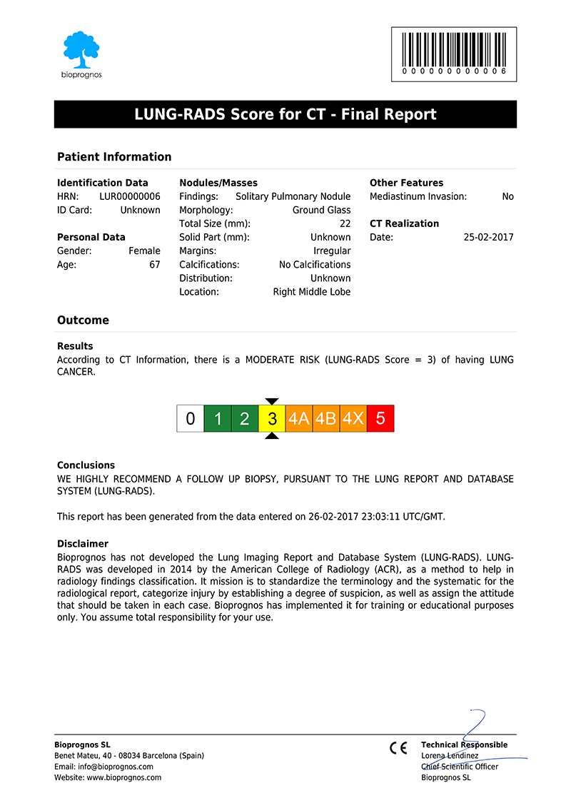
Lung Rads For Ct Scan Bioprognos
www.bioprognos.com
Decision Memo For Screening For Lung Cancer With Low Dose Computed Tomography Ldct Cag 00439n
www.cms.gov

Stages 2 3 And 4 Of Lung Cancer Download Scientific Diagram
www.researchgate.net

Small Cell Lung Carcinoma Staging Imaging And Treatment Considerations Radiographics
pubs.rsna.org

Slow Growing Lung Cancer As An Emerging Entity From Screening To Clinical Management European Respiratory Society
erj.ersjournals.com
Automatic Lung Nodule Detection Using Multi Scale Dot Nodule Enhancement Filter And Weighted Support Vector Machines In Chest Computed Tomography
journals.plos.org
Radiation For Lung Cancer
www.slideshare.net

Small Cell Lung Carcinoma Staging Imaging And Treatment Considerations Radiographics
pubs.rsna.org
Lung Cancer Wikipedia
en.wikipedia.org

Can Lung Cancer Be Missed On A Ct Scan Ct Scan Machine
ctscanmachines.blogspot.com
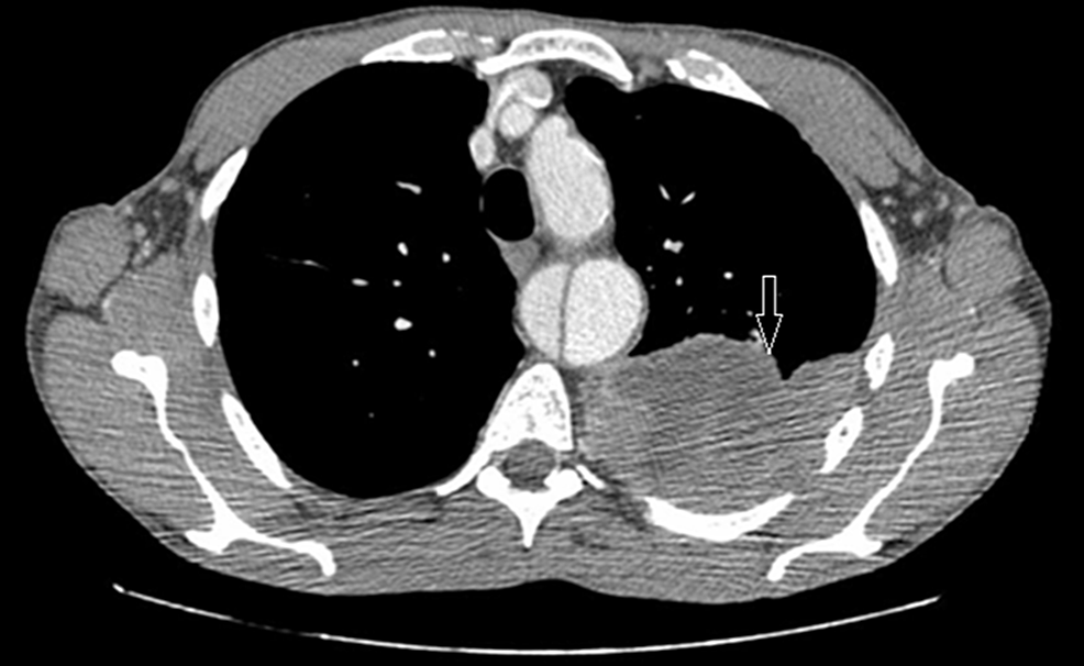
Cureus Spindle Cell Carcinoma Of The Lung Pleura An Incidental Finding
www.cureus.com
Decision Memo For Screening For Lung Cancer With Low Dose Computed Tomography Ldct Cag 00439n
www.cms.gov
Effective And Reliable Framework For Lung Nodules Detection From Ct Scan Images Scientific Reports
www.nature.com
Highly Accurate Model For Prediction Of Lung Nodule Malignancy With Ct Scans Scientific Reports
www.nature.com

Ct Databases Aimed At Lung Imaging Research
www.auntminnie.com
Slow Growing Lung Cancer As An Emerging Entity From Screening To Clinical Management European Respiratory Society
erj.ersjournals.com

Deep Learning Predicts Lung Cancer Treatment Response From Serial Medical Imaging Clinical Cancer Research
clincancerres.aacrjournals.org

Metastatic Nsclc Exploring New Therapeutic Combinations
www.medpagetoday.com

Standard Dose Vs Low Dose Ct Protocols In The Evaluation Of Localized Lung Lesions Capability For Lesion Characterization Ilead Study Sciencedirect
www.sciencedirect.com

Lung Cancer Screening Nodule Identification And Characterization Vlahos Translational Lung Cancer Research
tlcr.amegroups.com
Http Iopscience Iop Org Article 10 1088 1742 6596 893 1 012063 Pdf

Pdf Lung Cancer Diagnosis Using Ct Scan Images Based On Cellular Learning Automata
www.researchgate.net
2
Nih Clinical Center Provides One Of The Largest Publicly Available Chest X Ray Datasets To Scientific Community National Institutes Of Health Nih
www.nih.gov
A Resected Case Of Solitary Pancreatic Metastasis From Adenocarcinoma Of The Lung Insight Medical Publishing
pancreas.imedpub.com
Ct Scan Diagnosing Lung Cancer Cancer Research Uk
www.cancerresearchuk.org
Linear Atelectasis Around The Hilum On Chest Radiography A Novel Sign Of Early Lung Cancer Journal Of Clinical Imaging Science
clinicalimagingscience.org

Surgery For Nonsmall Cell Lung Cancer European Respiratory Society
err.ersjournals.com

Ct Medical Images Kaggle
www.kaggle.com

Medicare Will Pay For Lung Ct Scans For Cancer Screening
www.modernhealthcare.com
Lung Cancer Newsroom
news.weill.cornell.edu

Pdf Automatic 3d Pulmonary Nodule Detection In Ct Images A Survey
www.researchgate.net

Radiomics And Radiogenomics In Lung Cancer A Review For The Clinician Lung Cancer
www.lungcancerjournal.info

Computer Assisted Decision Support System In Pulmonary Cancer Detection And Stage Classification On Ct Images Sciencedirect
www.sciencedirect.com

Images Learn Nlst The Cancer Data Access System
cdas.cancer.gov

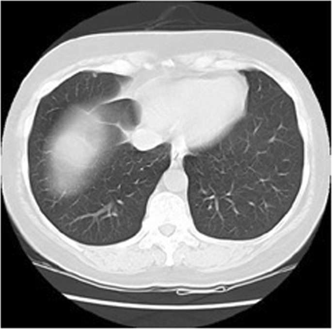
2d Ct Scan Slice Containing A Small 5mm Early Stage Lung Cancer Download Scientific Diagram
www.researchgate.net

Contribution Of Magnetic Resonance Imaging In Lung Cancer Imaging Sciencedirect
www.sciencedirect.com
Https Arxiv Org Pdf 1802 01756

Integrated Pet Ct In The Staging Of Nonsmall Cell Lung Cancer Technical Aspects And Clinical Integration European Respiratory Society
erj.ersjournals.com
Https Encrypted Tbn0 Gstatic Com Images Q Tbn 3aand9gcr5fgbmovb6ieimdpgjxowiaz0jgjollcg2zd90rdwzq S63so6 Usqp Cau
encrypted-tbn0.gstatic.com

Non Small Cell Lung Cancer Staging
imagingpathways.health.wa.gov.au
J Imaging Free Full Text Lung Nodule Detection In Ct Images Using Statistical And Shape Based Features Html
www.mdpi.com

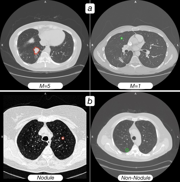
Pdf Cloud Based Nosql Open Database Of Pulmonary Nodules For Computer Aided Lung Cancer Diagnosis And Reproducible Research
www.researchgate.net
2
Decision Memo For Screening For Lung Cancer With Low Dose Computed Tomography Ldct Cag 00439n
www.cms.gov

A Computer Aided Pipeline For Automatic Lung Cancer Classification On Computed Tomography Scans
www.hindawi.com
