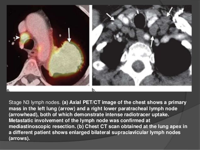
Lymph Node Lung Cancer Pet Ct Scan
Integrated Pet Ct In The Staging Of Nonsmall Cell Lung Cancer Technical Aspects And Clinical Integration European Respiratory Society
erj.ersjournals.com
Https Encrypted Tbn0 Gstatic Com Images Q Tbn 3aand9gcq6xjxerjxit4qxz Awgdqgljwdo9 5aocltsa6ftfkc5tdeu2q Usqp Cau
encrypted-tbn0.gstatic.com

Pet Ct Imaging In Lung Cancer Indications And Findings
www.scielo.br

Lung Cancer Snmmi
www.snmmi.org
The Value Of 18f Fdg Pet Ct Imaging In Breast Cancer Staging Bosnian Journal Of Basic Medical Sciences
www.bjbms.org
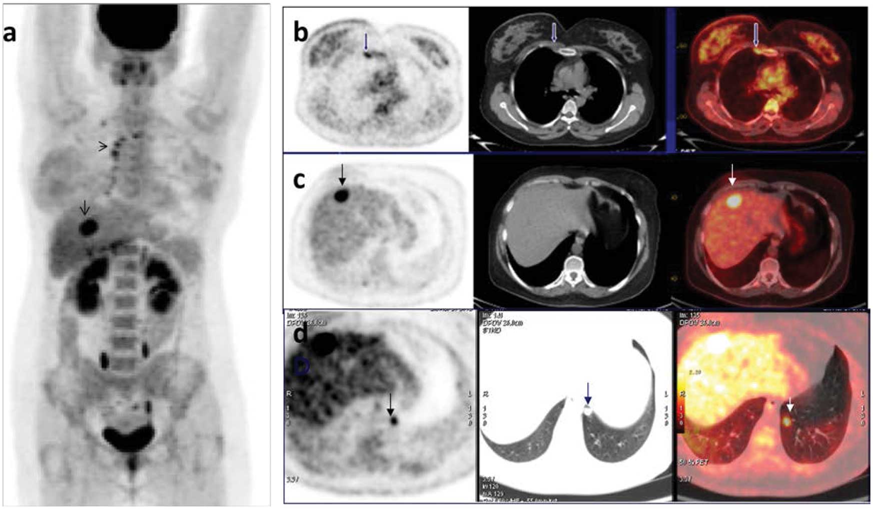
Coexistence Of Sarcoidosis And Metastatic Lesions A Diagnostic And Therapeutic Dilemma Review
www.spandidos-publications.com
A lung pet scan is typically combined with a lung ct scan to detect conditions like lung cancer.
Lymph node lung cancer pet ct scan. Ct scan showed slight increase in lymph node after 34 years of clean scans in. My wife was diagnosed in september 2007 with non small cell lung cancer with mets to the brain lymph nodes and adrenal glands. A ct scan takes a series of x rays that build up a three dimensional picture of the inside of the body.
This is the type of pet scan most often used in patients with lung cancer. To perform a prospective comparison of direct real time endobronchial ultrasound ebus guided transbronchial needle aspiration tbna positron emission tomography pet and thoracic ct for detection of mediastinal and hilar lymph node metastasis in patients with lung cancer considered for surgical resection. Petct scans can be useful.
How secondary cancer in the lymph nodes is diagnosed. The computer combines information from the two scans to provide a three dimensional image which. Often a pet scan is combined with a ct scan using a special machine that can do both at the same time.
The scan is painless and takes 10 15. Ct scan showed slight increase in lymph node after 34 years of clean scans in. Lung cancer survivors hello all i picked up my reports i was wondering if anyone else has had a similar scan report.
The ct scan only takes around two minutes. Sometimes a ct scan or mri scan is all that is needed to make a diagnosis of secondary cancer in the lymph nodes. Lung cancer survivors diagnosed with stage1b upper left lung adenocarcinoma january 2015.
If a pet ct scan is performed the ct scan will be performed first. The pet scan will follow and can take anywhere from 20 to 45 minutes depending on the purpose and scope of the test. It is part of a classification system called tnm staging which categorizes cancer by the size of the primary tumor t the number and location of regional lymph nodes n and the presence or absence of metastasis m.
Ct computerized tomography scan.
Prostate Pet Ct Targets More Cancer And Improves Patient Care Imaging Technology News
www.itnonline.com

Pdf Accuracy Of 18f Fdg Pet Ct For Lymph Node Staging In Non Small Cell Lung Cancers
www.researchgate.net

Pet Ct In The Assessment Of Lung Cancer At Rikshospitalet From 2007 2011 Tidsskrift For Den Norske Legeforening
tidsskriftet.no

Pet Ct Imaging In Lung Cancer Indications And Findings
www.scielo.br

Lung Cancer Combined 3d Ct And Pet Scans Stock Image C034 5669 Science Photo Library
www.sciencephoto.com

Pet Ct Imaging In Lung Cancer Indications And Findings
www.scielo.br
Usefulness Of Pet Ct In The Diagnosis Of Recurrent Or Metastasized Differentiated Thyroid Carcinoma
www.spandidos-publications.com
Frontiers Detection Of Metastatic Disease In Cardiophrenic Lymph Nodes Fdg Pet Ct Versus Contrast Enhanced Ct And Implications For Staging And Treatment Of Disease Oncology
www.frontiersin.org
Ct Pet Ct Mrt And Transthoracic Ultrasound In Lung Cancer Staging
healthcare-in-europe.com

Figure 1 From Pet In Lung Cancer Semantic Scholar
www.semanticscholar.org

Ct And Pet Lung Cancer
ykhoa.org

Radiologia Brasileira O Uso De Fdg Pet Tc Scan No Planejamento Da Radioterapia Em Cancer Do Pulmao Nao De Pequenas Celulas
www.rb.org.br
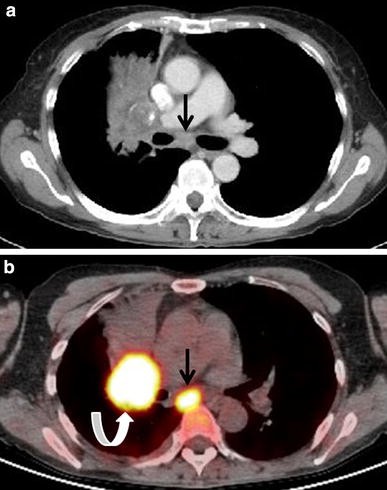
Figure 1 Present And Future Roles Of Fdg Pet Ct Imaging In The Management Of Lung Cancer Springerlink
link.springer.com

Xrays And Ct Scans Of Lung Cancer
www.aboutcancer.com

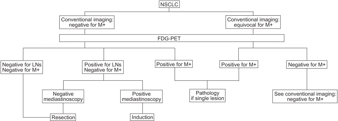
Algorithm For The Staging Of Nonsmall Cell Lung Cancer Nsclc With The Download Scientific Diagram
www.researchgate.net

Epos P 0031
epos.myesr.org
Psma Pet Ct Accurately Detects Prostate Cancer Spread National Cancer Institute
www.cancer.gov

Pet Ct Imaging In Lung Cancer Indications And Findings
www.scielo.br

Figure 3 From Fdg Pet Ct For Cancer Management Semantic Scholar
www.semanticscholar.org
Neoplasms Of The Lung Radiology Key
radiologykey.com
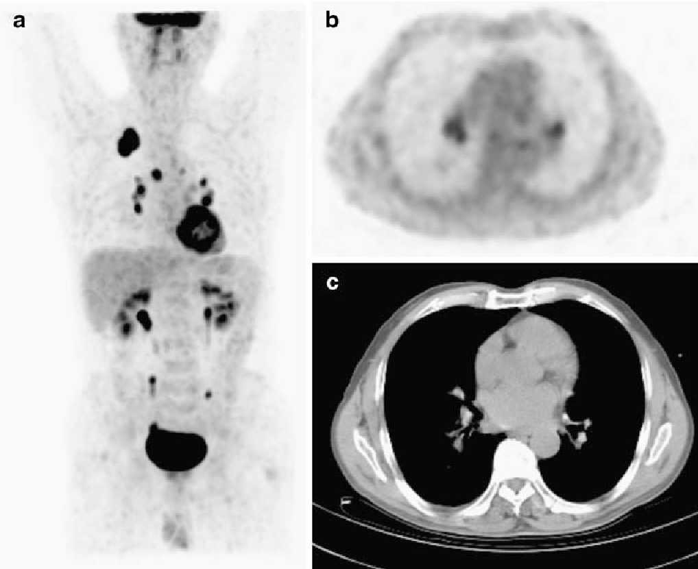
Figure 4 From 18f Fdg Pet Ct In Mediastinal Lymph Node Staging Of Non Small Cell Lung Cancer In A Tuberculosis Endemic Country Consideration Of Lymph Node Calcification And Distribution Pattern To Improve Specificity Semantic Scholar
www.semanticscholar.org

Pet Ct
www.med-ed.virginia.edu

Preoperative Staging Of Lung Cancer With Combined Pet Ct Nejm
www.nejm.org

Integrated Pet Ct In The Staging Of Nonsmall Cell Lung Cancer Technical Aspects And Clinical Integration European Respiratory Society
erj.ersjournals.com

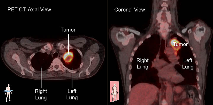
Pet Ct Scan Shows One Pulmonary Nodule At The Left Upper Lobe With Download Scientific Diagram
www.researchgate.net

18f Fdg Pet Ct Imaging Of The Pancreas Spectrum Of Diseases Insight Medical Publishing
pancreas.imedpub.com

The Radiology Assistant Tnm Classification 8th Edition
radiologyassistant.nl

Pet Ct
www.med-ed.virginia.edu

Xrays And Ct Scans Of Lung Cancer
www.aboutcancer.com

Figure 1 From The Diagnostic Contribution Of 18 F Fdg Pet Ct Scan In Cancer Of Unknown Primary Semantic Scholar
www.semanticscholar.org
Imaging Tests Lungevity Foundation
lungevity.org
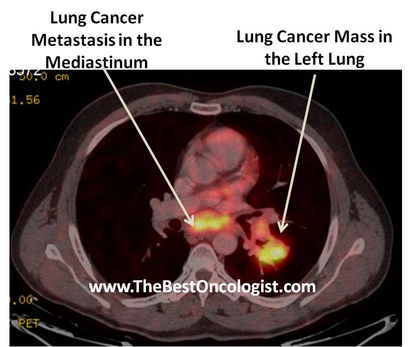
How Lung Cancer Is Diagnosed The Best Oncologist Tm
www.thebestoncologist.com

Staging Of Non Small Cell Lung Cancer With Integrated Positron Emission Tomography And Computed Tomography Nejm
www.nejm.org
Quantitative Fdg Pet Ct May Help Risk Stratify Early Stage Non Small Cell Lung Cancer Patients At Risk For Recurrence Following Anatomic Resection Harmon Journal Of Thoracic Disease
jtd.amegroups.com

Diagnostic Accuracy Of Virtual 18f Fdg Pet Ct Bronchoscopy For The Detection Of Lymph Node Metastases In Non Small Cell Lung Cancer Patients
jnm.snmjournals.org
Lung Cancer Radiology
www.slideshare.net

Thoracic Lymph Node Stations Annotated Ct Radiology Case Radiopaedia Org
radiopaedia.org

Pet Ct In Nononcological Lung Diseases Current Applications And Future Perspectives European Respiratory Society
err.ersjournals.com
Plos One A Decision Tree Model For Predicting Mediastinal Lymph Node Metastasis In Non Small Cell Lung Cancer With F 18 Fdg Pet Ct
journals.plos.org

Pet Ct Open Air Mri Of Cen La
openairmri.com

Pet Ct
www.med-ed.virginia.edu
Pet Ct Principles And Applications In Lung Cancer Management Intechopen
www.intechopen.com
Xrays And Ct Scans Of Lung Cancer
www.aboutcancer.com

Lung Cancer Metastasis To Subcarinal Level 7 Lymph Node Although Download Scientific Diagram
www.researchgate.net
Plos One Correlation Of The Apparent Diffusion Coefficient Adc With The Standardized Uptake Value Suv In Lymph Node Metastases Of Non Small Cell Lung Cancer Nsclc Patients Using Hybrid 18f Fdg Pet Mri
journals.plos.org
Pet Ct Principles And Applications In Lung Cancer Management Intechopen
www.intechopen.com

Imaging Of Lung Cancer Implications On Staging And Management Purandare Nc Rangarajan V Indian J Radiol Imaging
www.ijri.org

Pet Ct Imaging For Target Volume Delineation In Curative Intent Radiotherapy Of Non Small Cell Lung Cancer Iaea Consensus Report 2014 Radiotherapy And Oncology
www.thegreenjournal.com

Lung Cancer Metastasis To Subcarinal Level 7 Lymph Node Although Download Scientific Diagram
www.researchgate.net
Integrated Pet Ct In The Staging Of Nonsmall Cell Lung Cancer Technical Aspects And Clinical Integration European Respiratory Society
erj.ersjournals.com

Mediastinal Lymph Node Staging Of Non Small Cell Lung Cancer A Prospective Comparison Of Computed Tomography And Positron Emission Tomography Sciencedirect
www.sciencedirect.com
Figure 1 From The Role Of 18 F Fdg Pet Ct For Evaluation Of Metastatic Mediastinal Lymph Nodes In Patients With Lung Squamous Cell Carcinoma Or Adenocarcinoma Semantic Scholar
www.semanticscholar.org
Isolated Supraclavicular Lymph Node Metastasis In Pancreatic Adenocarcinoma A Report Of Three Cases And Review Of The Literature Insight Medical Publishing
pancreas.imedpub.com
Retrospective Comparative Study Between 3t Wb Mri Including Wb Dwi And 18f Fdg Pet Ct In Detection Of Metastatic Disease
www.oatext.com
Surgical Resection And Long Term Disease Free Survival In Stage Iiib Non Small Cell Lung Cancer After Gefitinib Down Staging A Case Report
www.oatext.com

Preoperative Staging Of Non Small Cell Lung Cancer With Positron Emission Tomography Nejm
www.nejm.org

Fdg Pet Ct For Solitary Pulmonary Nodule And Lung Cancer Literature Review Sciencedirect
www.sciencedirect.com

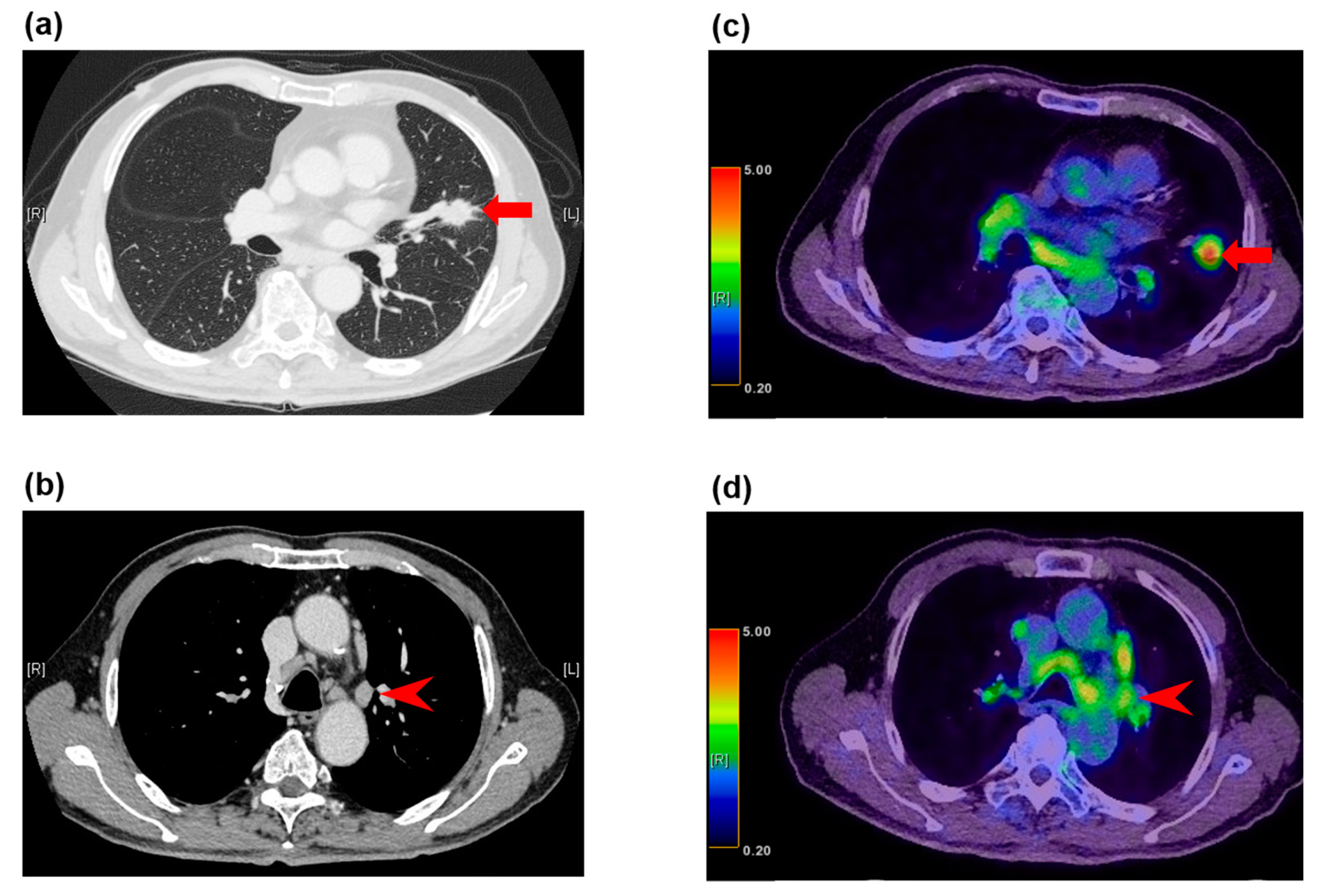
A Representative Case Of 18 F Fdg Pet Ct For Lymph Node Staging In Lung Download Scientific Diagram
www.researchgate.net
Present And Future Roles Of Fdg Pet Ct Imaging In The Management Of Lung Cancer Springerlink
link.springer.com

Clinical Utility Of Pet Scanning In Breast Cancer Oncology Cme
www.gotoper.com

Lung Cancer Ct And Pet Scan Stock Image C016 6772 Science Photo Library
www.sciencephoto.com
Improved Detection Of Metastatic Lymph Nodes In Oesophageal Squamous Cell Carcinoma By Combined Interpretation Of Fluorine 18 Fluorodeoxyglucose Positron Emission Tomography Computed Tomography Cancer Imaging Full Text
cancerimagingjournal.biomedcentral.com
Revisions To The Tumor Node Metastasis Staging Of Lung Cancer 8th Edition Rationale Radiologic Findings And Clinical Implications
www.wjgnet.com
Clinical Impact Of 18f Fdg Pet Ct On Initial Staging And Therapy Planning For Breast Cancer
www.spandidos-publications.com
Pet Ct Imaging Pre Chemotherapy A The Coronal 18fdg Pet Ct Scans Download Scientific Diagram
www.researchgate.net

Preoperative Staging Of Non Small Cell Lung Cancer With Positron Emission Tomography Nejm
www.nejm.org
Updates On 18 F Fdg Pet Ct As A Clinical Tool For Tuberculosis Evaluation And Therapeutic Monitoring Yu Quantitative Imaging In Medicine And Surgery
qims.amegroups.com

Invasive Mediastinal Staging Recommendations This Algorithm Applies To Download Scientific Diagram
www.researchgate.net

Common Causes Of False Positive F18 Fdg Pet Ct Scans In Oncology
www.scielo.br

Diagnosis And Staging Of Lung Cancer Disease Any Disease S Symptoms
anydiseasesymptoms.com
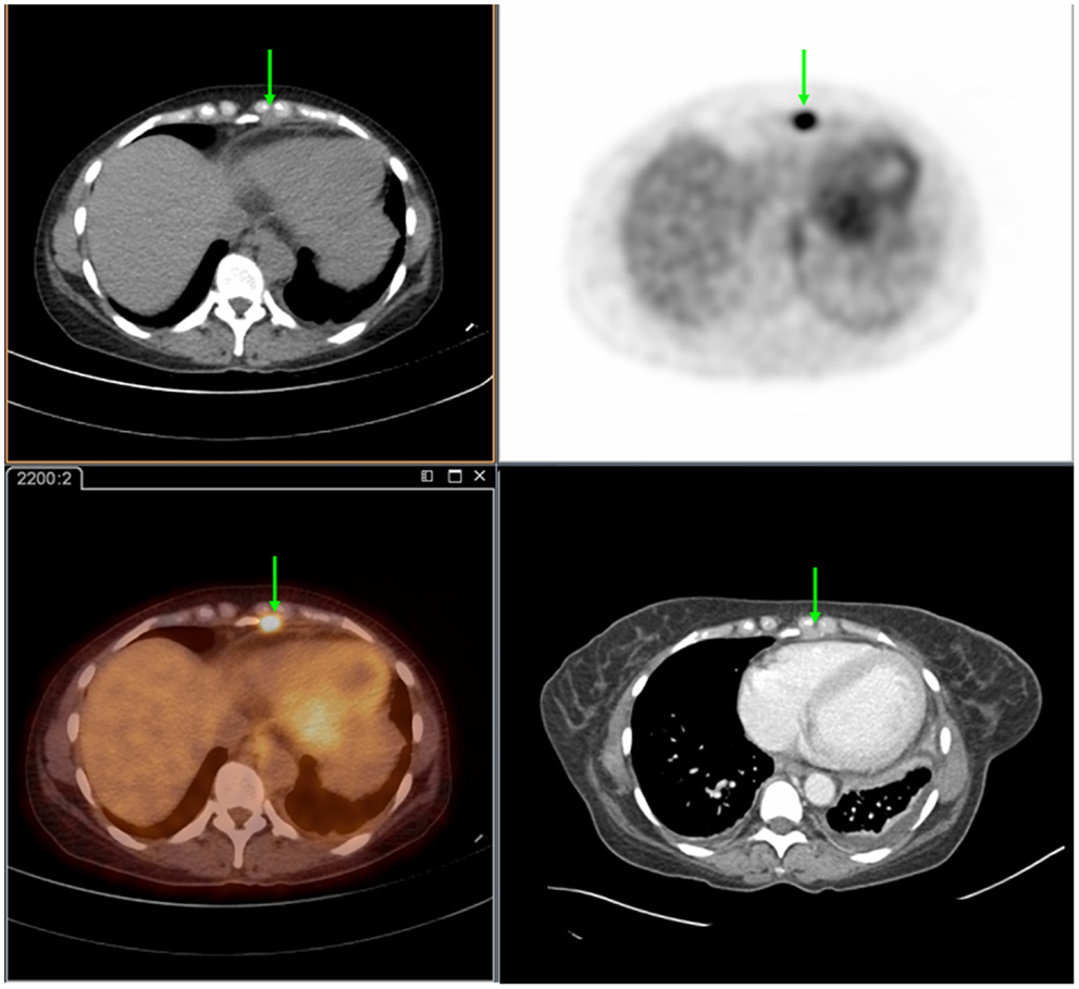
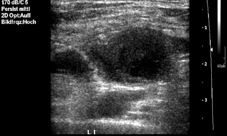
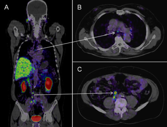
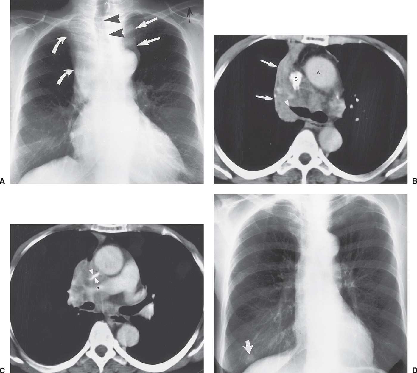
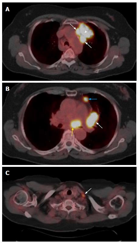
Case Report
pulmonarychronicles.com

Ct Imaging In Pre Therapeutic Assessment Of Lung Cancer Sciencedirect
www.sciencedirect.com

Pet Ct In Anal Cancer Indications And Limits Intechopen
www.intechopen.com
Diagnostics Free Full Text Recent And Current Advances In Fdg Pet Imaging Within The Field Of Clinical Oncology In Nsclc A Review Of The Literature Html
www.mdpi.com

Xrays And Ct Scans Of Lung Cancer
www.aboutcancer.com

Role Of Positron Emission Tomography Computed Tomography In Screening Metastasis Of Renal Cell Carcinoma Sciencedirect
www.sciencedirect.com

Preoperative Staging Of Non Small Cell Lung Cancer With Positron Emission Tomography Nejm
www.nejm.org
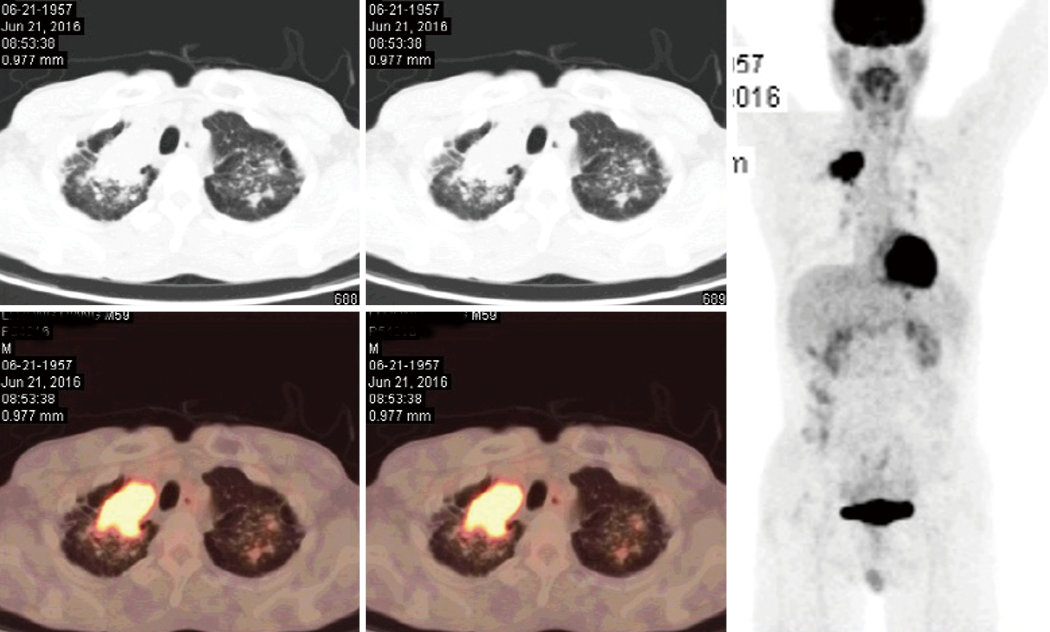
A Case Of Primary Lung Cancer Lesion Demonstrated By F 18 Fdg Positron Emission Tomography Computed Tomography Pet Ct One Year After The Detection Of Metastatic Brain Tumor
www.spandidos-publications.com
Https Encrypted Tbn0 Gstatic Com Images Q Tbn 3aand9gcqscm8qlx9qwapcwilybirptagvuhn1elwhse0vz1jsa Jtsfgq Usqp Cau
encrypted-tbn0.gstatic.com
:max_bytes(150000):strip_icc()/lung-cancer-spread-to-lymph-nodes-2249364-v2-bef5b796e83a46ceb3a50d82ca1c71e3.png)
When Lung Cancer Spreads To Lymph Nodes
www.verywellhealth.com
Frontiers Current Concepts In F18 Fdg Pet Ct Based Radiation Therapy Planning For Lung Cancer Oncology
www.frontiersin.org

Early And Locally Advanced Non Small Cell Lung Cancer Nsclc Esmo Clinical Practice Guidelines For Diagnosis Treatment And Follow Up Annals Of Oncology
www.annalsofoncology.org
Table 1 From 18f Fdg Pet Ct In Mediastinal Lymph Node Staging Of Non Small Cell Lung Cancer In A Tuberculosis Endemic Country Consideration Of Lymph Node Calcification And Distribution Pattern To Improve Specificity Semantic Scholar
www.semanticscholar.org

Figure 1 From Current Concepts In F18 Fdg Pet Ct Based Radiation Therapy Planning For Lung Cancer Semantic Scholar
www.semanticscholar.org

A Representative Case Of 18 F Fdg Pet Ct For Lymph Node Staging In Lung Download Scientific Diagram
www.researchgate.net
Https Encrypted Tbn0 Gstatic Com Images Q Tbn 3aand9gctbt6avfwdwc7 Kcxtlsd5blbqzxqeqszgp2olleszshofk78y0 Usqp Cau
encrypted-tbn0.gstatic.com

Pet Ct 1 Year After Pulmonary Surgery Showing Massive F Open I
openi.nlm.nih.gov

Pet Ct Imaging In Lung Cancer Indications And Findings
www.scielo.br
1
encrypted-tbn0.gstatic.com
Plos One Correlation Of The Apparent Diffusion Coefficient Adc With The Standardized Uptake Value Suv In Lymph Node Metastases Of Non Small Cell Lung Cancer Nsclc Patients Using Hybrid 18f Fdg Pet Mri
journals.plos.org

Fdg Pet Ct For Solitary Pulmonary Nodule And Lung Cancer Literature Review Sciencedirect
www.sciencedirect.com

Xrays And Ct Scans Of Lung Cancer
www.aboutcancer.com

Fdg Pet Ct For Solitary Pulmonary Nodule And Lung Cancer Literature Review Sciencedirect
www.sciencedirect.com
Diagnostic Performance Of Integrated Positron Emission Tomography Computed Tomography For Mediastinal Lymph Node Staging In Non Small Cell Lung Cancer Ppt Download
slideplayer.com
Present And Future Roles Of Fdg Pet Ct Imaging In The Management Of Lung Cancer Springerlink
link.springer.com

18f Fdg Pet Ct Imaging Of The Pancreas Spectrum Of Diseases Insight Medical Publishing
pancreas.imedpub.com

Small Cell Lung Cancer Chest Ct Scan A And Corresponding Pet Ct Download Scientific Diagram
www.researchgate.net

A Pet Ct Scan Demonstrating A Left Hilar Lymph Node With Positivity Download Scientific Diagram
www.researchgate.net
