Pet Ct Esophageal Cancer

Esophageal Cancer With Positive Nodes On The Pet Ct And Ct Esophagus Case Studies Ctisus Ct Scanning
ctisus.com

Pet Ct Open Air Mri Of Cen La
openairmri.com
Esophageal Cancer Radiology Key
radiologykey.com

Fdg Pet Ct Has Limits In Esophageal Cancer Follow Up
www.auntminnieeurope.com

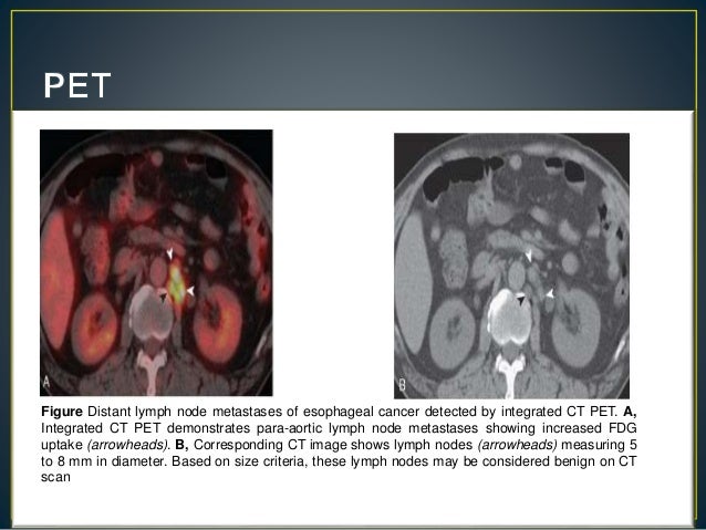
Pet Ct Of Esophageal Cancer Its Role In Clinical Management Radiographics
pubs.rsna.org

Cxcr4 Is A Potential Target For Diagnostic Pet Ct Imaging In Barrett S Dysplasia And Esophageal Adenocarcinoma Clinical Cancer Research
clincancerres.aacrjournals.org
This is a bibliography of relevent articles on petct for esophagus cancer.

Pet ct esophageal cancer. Am j clin pathol 2012137516 542. You have a pet ct scan to find out more about where exactly your oesophageal cancer is and whether it has spread. If these tests do not clearly show that an abnormality is benign a biopsy may be necessary.
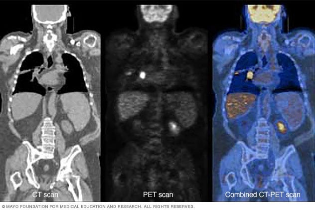
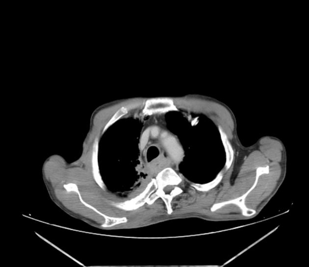
At left is a ct scan while the center image is from a pet scanner. This test for esophageal cancer is designed to detect abnormalities in the surrounding tissue and lymph nodes. A combination of ct scan transesophageal ultrasound and petct scan are used for staging of the disease.
Ct scan for esophageal cancer uses x ray images to present detailed images of the esophagus and surrounding tissues. Sometimes a pet scan is combined with a ct scan using a special machine that can do both at the same time. American cancer society american society for colposcopy and cervical pathology and american society for clinical pathology screening guidelines for the prevention and early detection of cervical cancer.
It also helps. In diagnosing esophageal cancer. Esophageal cancer is often treated by using a combined modality approach chemotherapy radiation therapy and esophagectomy.
Grilo a vieira l carolino e et al. An eus also helps stage esophageal cancer which guides treatment decisions and prognosis assessments. It gives detailed information.
Petct can detect esophageal cancer determine if it has spread assess the effectiveness of a treatment plan and determine if the cancer has returned after treatment. Combining a pet scan with an mri or ct scan can help make the images easier to interpret. A positron emission tomography pet scan detects abnormal cell metabolism to diagnose cancer heart disease and brain disorders before other tests can.
A biopsy is the removal of tissue in order to examine it for disease. So petct is a relatively new very powerful imaging methodology for the diagnosis and staging of a wide range of cancers including esophageal cancer. It can help doctors work out whether tissue is active cancer or not.
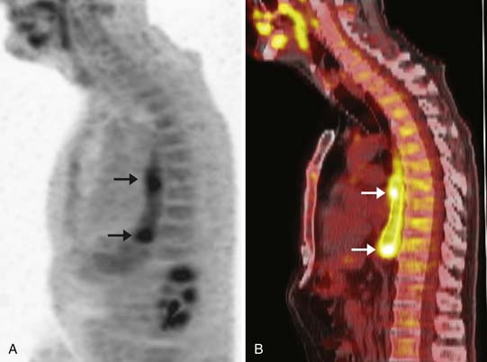
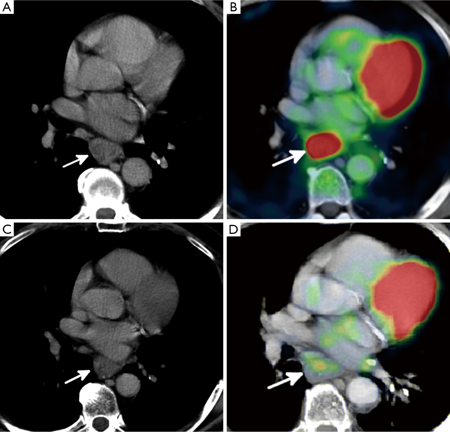
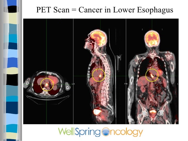
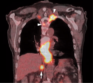
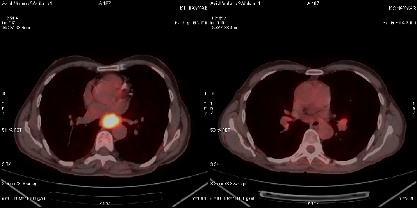
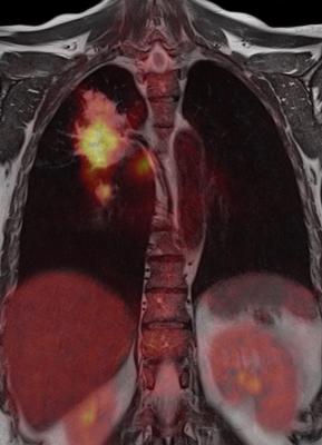
The bright spot in the chest seen best on the pet and ct pet scans is lung cancer. Fdg petct is a whole body technique with good accuracy for distant metastatic disease. A comparison of anxiety levels.
Correct integration of petct into the conventional work up of esophageal cancer requires a multidisciplinary approach that combines the information from petct with results of clinical assessment diagnostic ct. For the prevention and early detection of cervical cancer. Petct scans can be useful.
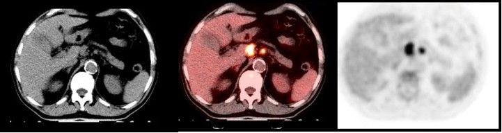
What is a pet ct scan. A pet ct scan combines a ct scan and a pet scan. This lets the doctor compare areas of higher radioactivity on the pet scan with the more detailed picture of that area on the ct scan.
Lung liver adrenal glands. Esophageal cancer colorectal cancer lymphoma melanoma. Anxiety in cancer patients during f fdg petct low dose.
1
encrypted-tbn0.gstatic.com

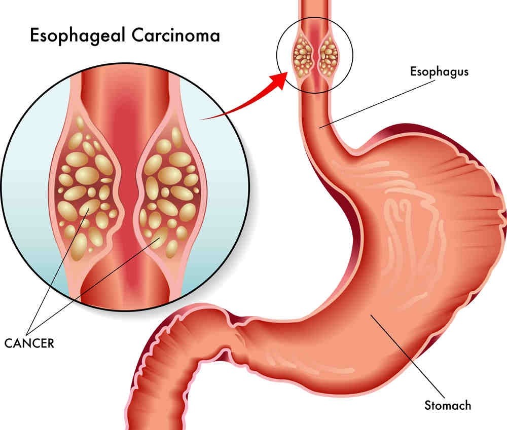
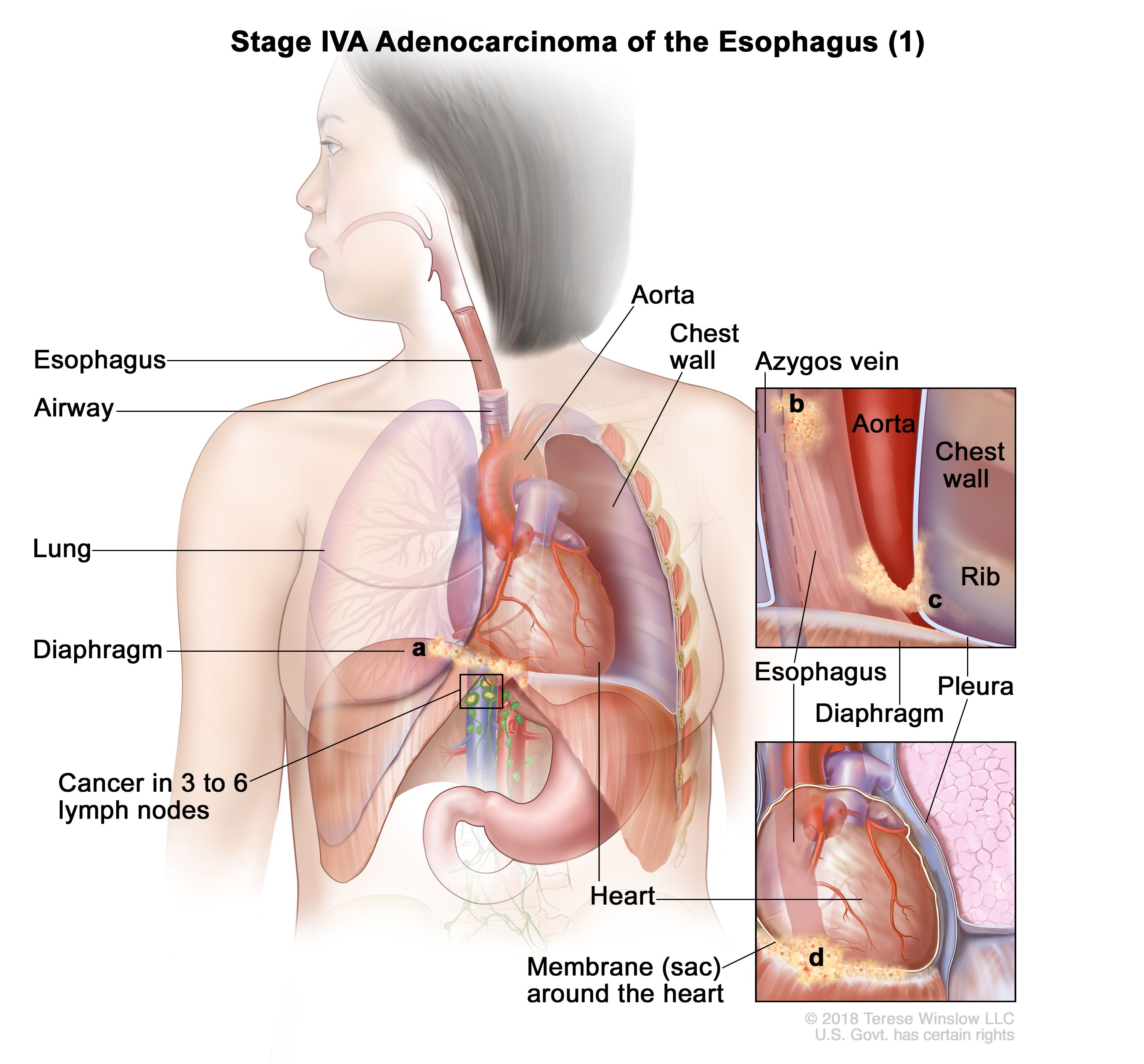
Anatomy Of The Esophagus
www.aboutcancer.com

Fluorodeoxyglucose Positron Emission Tomography Imaging Of Head And Neck Squamous Cell Cancer American Journal Of Neuroradiology
www.ajnr.org
Gastric Esophageal And Gastrointestinal Stromal Tumors Radiology Key
radiologykey.com
Role Of Fused Pet Ct Compared To The Standard Contrast Enhanced Ct In The Follow Up Assessment Of The Treated Gastric Malignancy Egyptian Journal Of Radiology And Nuclear Medicine Full Text
ejrnm.springeropen.com

Esophageal Cancer Diagnosis Imaging Tests Moffitt
moffitt.org
Positron Emission Tomography Scan Mayo Clinic
www.mayoclinic.org
State Of The Art Molecular Imaging In Esophageal Cancer Management Implications For Diagnosis Prognosis And Treatment Lin Journal Of Gastrointestinal Oncology
jgo.amegroups.com

Pet Ct
www.med-ed.virginia.edu

Pet Ct Planning During Chemoradiotherapy For Esophageal Cancer
www.e-roj.org
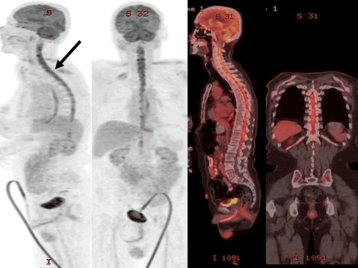
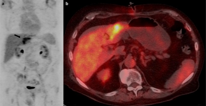
Intramedullary Metastases Detected On 18f Fdg Pet Ct Imaging
www.oatext.com

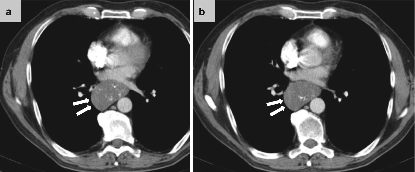
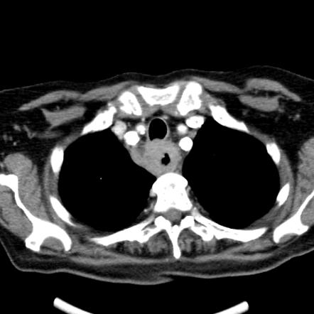
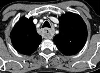
A 75 Year Old Man With Biopsy Proven Esophageal Cancer A Axial Ct Download Scientific Diagram
www.researchgate.net

Esophageal Cancer Devastating And Deadly
reference.medscape.com

Fdg Pet Ct Can Provide Valuable Esophageal Cancer Updates
www.auntminnieeurope.com

Overview Of The Tumor Delineation For One Patient With Middle Thoracic Download Scientific Diagram
www.researchgate.net
Carcinoma Of Esophagus
www.slideshare.net

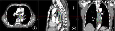
Esophageal Cancer With Positive Nodes On The Pet Ct And Ct Esophagus Case Studies Ctisus Ct Scanning
www.ctisus.com
Https Pubs Rsna Org Doi Pdf 10 1148 Rg 276065742
Esophageal Cancer Frequently Asked Questions
www.laparoscopyhospital.com
Comparison Of Gross Target Volumes Based On Four Dimensional Ct Positron Emission Tomography Computed Tomography And Magnetic Resonance Imaging In Thoracic Esophageal Cancer Cancer Med X Mol
www.x-mol.com

Appropriateness Criteria Of Fdg Pet Ct In Oncology Agrawal A Rangarajan V Indian J Radiol Imaging
www.ijri.org
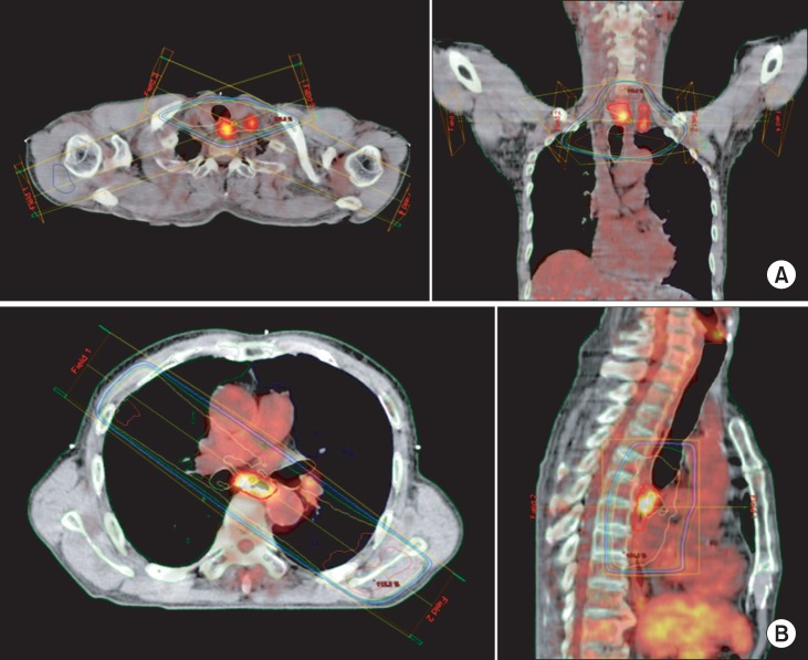
Pet Ct Planning During Chemoradiotherapy For Esophageal Cancer
www.e-roj.org

Anatomy Of The Esophagus
www.aboutcancer.com
Imaging Of The Stomach And Esophagus Using Ct And Pet Ct Techniques Springerlink
link.springer.com
Esophageal Cancer Pet Scans Are Key To Accurate Staging Peoplebeatingcancer
peoplebeatingcancer.org

Esophageal Cancer J Timothy Sherwood Md Thoracic Surgeon Ppt Video Online Download
slideplayer.com
Plos One 18f Fdg Pet Ct After Neoadjuvant Chemoradiotherapy In Esophageal Cancer Patients To Optimize Surgical Decision Making
journals.plos.org

Case Of The Quarter April 2011
www.nepetimaging.com
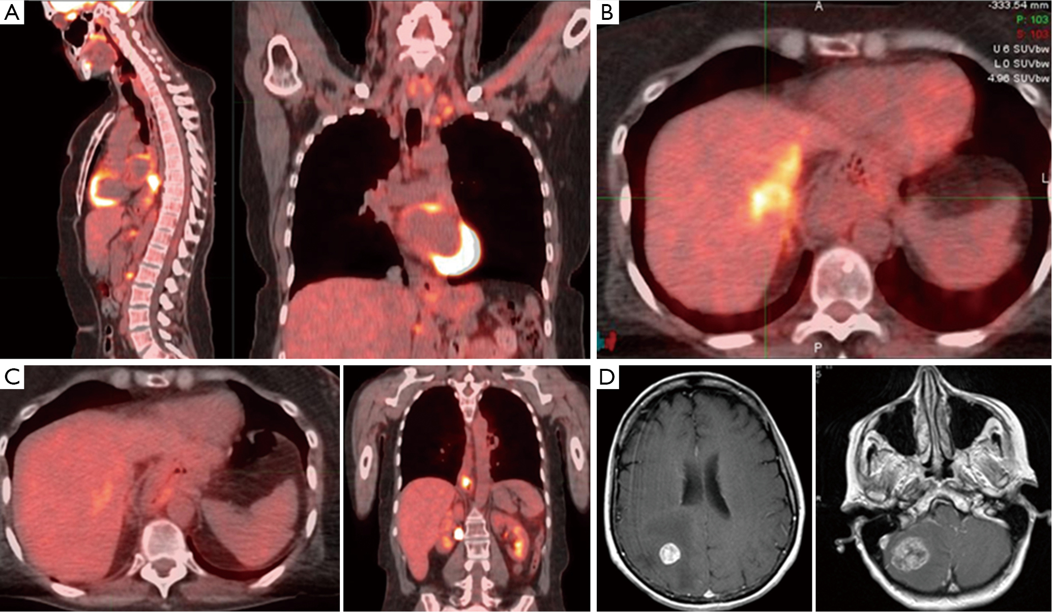
Her2 Positive Trastuzumab Resistant Metastatic Esophageal Cancer Presenting With Brain Metastasis After Durable Response To Dual Her2 Blockade A Case Report Niu Journal Of Gastrointestinal Oncology
jgo.amegroups.com
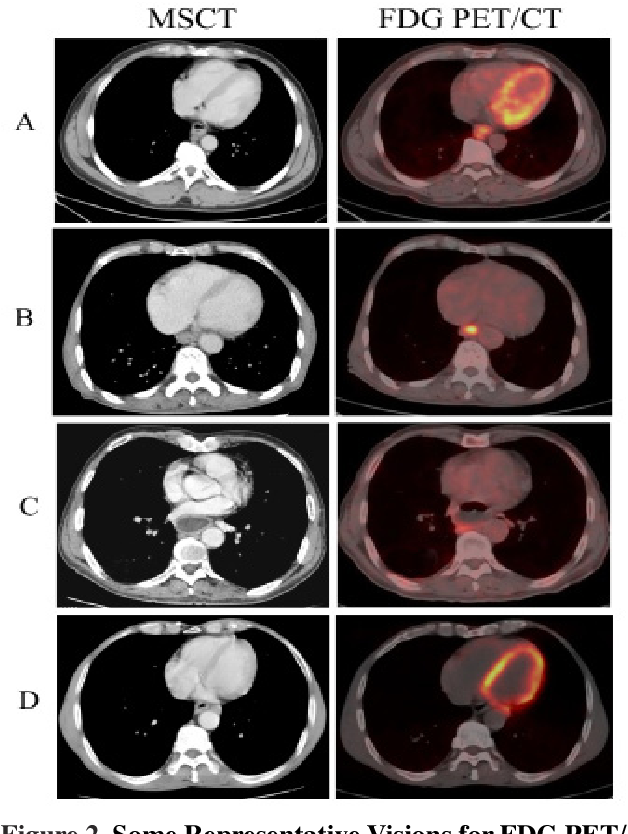
Figure 2 From Combination Of Fdg Pet Ct And Contrast Enhanced Msct In Detecting Lymph Node Metastasis Of Esophageal Cancer Semantic Scholar
www.semanticscholar.org

Coexisting Laryngeal Tumor In A Patient With Esophageal Carcinoma Successfully Treated With 18f Fdg Pet Ct Based Radiation Therapy Siemens Healthineers Slovakia
www.siemens-healthineers.com

18f Fdg Pet And 18f Fdg Pet Ct For Assessing Response To Therapy In Esophageal Cancer
jnm.snmjournals.org
Efficacy Of 4 Methyl 11c Thiothymidine Pet Ct Before And After Neoadjuvant Therapy For Predicting Therapeutic Responses In Patients With Esophageal Cancer A Pilot Study Ejnmmi Research Full Text
ejnmmires.springeropen.com
Figure 3 From Successfully Treated Advanced Esophageal Cancer With Left Axillary Lymph Node Metastasis And Synchronous Right Breast Cancer A Case Report Semantic Scholar
www.semanticscholar.org
Esophageal Carcinoma Radiology Reference Article Radiopaedia Org
radiopaedia.org

Pet Mri Holds Its Own For Esophageal Cancer Staging
www.auntminnie.com

Treatment Of A Solitary Cervical Lymph Node Recurrence Following Curative Resection For Thoracic Oesophageal Cancer Digitale Posterprasentation
sgc2014.kongress-poster.ch
Is Endoscopic Ultrasound Examination Necessary In The Management Of Esophageal Cancer
www.wjgnet.com
Esophagus
www.slideshare.net

Pet Ct
www.med-ed.virginia.edu

Cancer Health Basics Esophageal Cancer Cancer Health
www.cancerhealth.com

Pet Ct Esophageal Cancer
www.cedars-sinai.edu

Esophageal Cancer Ppt Video Online Download
slideplayer.com
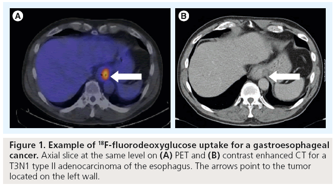
Image Guided Radiotherapy For Esophageal Cancer
www.openaccessjournals.com

Coexisting Laryngeal Tumor In A Patient With Esophageal Carcinoma Successfully Treated With 18f Fdg Pet Ct Based Radiation Therapy Siemens Healthineers Slovakia
www.siemens-healthineers.com

Xmlinkhub
jkms.org

Positron Emission Tomography Pet Special Subjects Msd Manual Consumer Version
www.msdmanuals.com

Pdf Pet Ct Of Esophageal Cancer Its Role In Clinical Management
www.researchgate.net
Pet Ct Praxis Fur Nuklearmedizin Pet Ct Zentrum
www.curanosticum.de

Role Of Fluorine 18 Fluorodeoxyglucose Positron Emission Tomography Computed Tomography In Gastrointestinal Cancers Sciencedirect
www.sciencedirect.com
Gastric Esophageal And Gastrointestinal Stromal Tumors Radiology Key
radiologykey.com

Pitfalls And Artifacts In The Interpretation Of Oncologic Pet Ct Of The Chest
www.scielo.br

Pdf Pet And Pet Ct Imaging In Esophageal And Gastric Cancers Semantic Scholar
www.semanticscholar.org
18f Fluorodeoxyglucose Positron Emission Computed Tomography For Monitoring Tumor Response In Esophageal Carcinoma Treated With Concurrent Chemoradiotherapy
www.spandidos-publications.com
Metastatic Esophageal Squamous Cell Carcinoma Successfully Treated With Immunotherapy First Case Report In Thailand
dx.doi.org
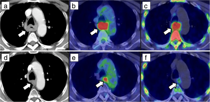
Improved Detection Of Metastatic Lymph Nodes In Oesophageal Squamous Cell Carcinoma By Combined Interpretation Of Fluorine 18 Fluorodeoxyglucose Positron Emission Tomography Computed Tomography Cancer Imaging Full Text
cancerimagingjournal.biomedcentral.com
Esophageal Cancer With Pulmonary Metastasis Radiology Case Radiopaedia Org
radiopaedia.org
Ssat Pet Ct For Response Assessment Of Neoadjuvant Chemoradiation In Locally Advanced Squamous Cell Carcinoma Esophagus Initial Experience From Tertiary Referral Center In North India
meetings.ssat.com
What Are The Most Common Esophageal Metastases
www.cancernetwork.com
Clinical Impact Of Fdg Pet Ct In Alimentary Tract Malignancies An Updated Review Springerlink
link.springer.com
Study Suggests Lung Cancer Survivors Receiving Too Many Pet Scans Imaging Technology News
www.itnonline.com

Pitfalls And Artifacts In The Interpretation Of Oncologic Pet Ct Of The Chest
www.scielo.br

Positron Emission Tomography Computed Tomography Pet Ct Findings A Download Scientific Diagram
www.researchgate.net

Anatomy Of The Esophagus
www.aboutcancer.com

A Malignant Birthmark Detected By 18 F Fdg Pet Ct In A Patient With Esophageal Cancer Springerlink
www.x-mol.com
Fluorodeoxyglucose Positron Emission Tomography Imaging Of Head And Neck Squamous Cell Cancer American Journal Of Neuroradiology
www.ajnr.org

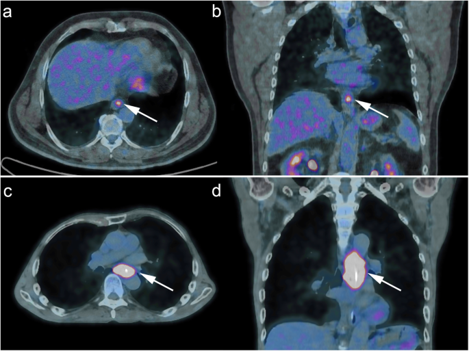
Fdg Pet Ct Image Demonstrating Increased Uptake At The Distal Download Scientific Diagram
www.researchgate.net
18f Fdg Pet And 18f Fdg Pet Ct For Assessing Response To Therapy In Esophageal Cancer
jnm.snmjournals.org

Pet Positive Esophageal Leiomyoma A Case Series And Literature Review Sages Abstract Archives
www.sages.org

Surgically Resected T1 And T2 Stage Esophageal Squamous Cell Carcinoma T And N Staging Performance Of Eus And Pet Ct Jeong 2018 Cancer Medicine Wiley Online Library
onlinelibrary.wiley.com

A 56 Year Old Man Who Came For Initial Staging Of Esophageal Cancer Download Scientific Diagram
www.researchgate.net

Use Of Pretreatment Metabolic Tumor Volumes On Pet Ct To Predict The Survival Of Patients With Squamous Cell Carcinoma Of Esophagus Treated By Curative Surgery
ar.iiarjournals.org
Outcomes Of Aggressive Treatment In Esophageal Cancer Patients With Synchronous Solitary Brain Metastasis
www.spandidos-publications.com
Diagnostics Free Full Text Clinical Utility Of Positron Emission Tomography Magnetic Resonance Imaging Pet Mri In Gastrointestinal Cancers Html
www.mdpi.com

Pitfalls And Artifacts In The Interpretation Of Oncologic Pet Ct Of The Chest
www.scielo.br
State Of The Art Molecular Imaging In Esophageal Cancer Management Implications For Diagnosis Prognosis And Treatment Lin Journal Of Gastrointestinal Oncology
jgo.amegroups.com
Pet Ct In Oncology Rcp Journals
www.rcpjournals.org

Positron Emission Tomography Pet Snmmi
www.snmmi.org

Esophageal Cancer Lakeshore Cancer Center
www.lakeshorecancercenter.org
Figure 1 From Clinical Implication Of Pet Mr Imaging In Preoperative Esophageal Cancer Staging Comparison With Pet Ct Endoscopic Ultrasonography And Ct Semantic Scholar
www.semanticscholar.org

Pet Ct For Detecting Recurrent Lymphoma
www.nepetimaging.com

Esophageal Cancer Symptoms Treatment Causes Prognosis
www.medicinenet.com
Esophageal Cancer Treatment Adult Pdq Health Professional Version National Cancer Institute
www.cancer.gov
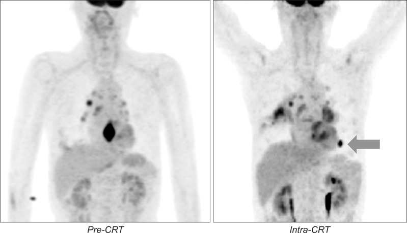
Pet Ct Planning During Chemoradiotherapy For Esophageal Cancer
www.e-roj.org

Medpix Case Squamous Cell Esophageal Cancer
medpix.nlm.nih.gov
Https Encrypted Tbn0 Gstatic Com Images Q Tbn 3aand9gcssay36lb3fqob8tn01u2 Pkzab9y3uceewzzevdgoxopjcuzbg Usqp Cau
encrypted-tbn0.gstatic.com
Pet Mr Could Be Effective For Staging Esophageal Cancer
www.cancernetwork.com

Quintessential Pet Scan Does Not Detect Cancer Cells
macsforcancer.com

Pet In Esophageal Cancer
www.auntminnie.com
18 F Fdg Pet Ct Derived Parameters Predict Clinical Stage And Prognosis Of Esophageal Cancer Bmc Medical Imaging Full Text
bmcmedimaging.biomedcentral.com
State Of The Art Molecular Imaging In Esophageal Cancer Management Implications For Diagnosis Prognosis And Treatment Lin Journal Of Gastrointestinal Oncology
jgo.amegroups.com

Chest X Ray And Pet Ct Findings Of A Patient With Advanced Esophageal Download Scientific Diagram
www.researchgate.net

Pet Ct Of Esophageal Cancer Its Role In Clinical Management Radiographics
pubs.rsna.org

Esophageal Carcinoma Current Concepts In The Role Of Imaging In Staging And Management Sciencedirect
www.sciencedirect.com

Pet Ct For Esophageal Carcinoma Staging Detects Clinically Unsuspected Synchronous Malignancy
www.healio.com

Pet Ct
www.med-ed.virginia.edu
