Pet Ct Scan For Liver Cancer

Pet Ct Scan Vs Ct Scan For Cancer Diagnosis
www.ctoam.com
What Is Pet Imaging Imaging Technology News
www.itnonline.com

First Total Body Pet Ct Studies Show Potential For Better Faster Lower Dose Images
medicalxpress.com
Exported Atypical Liver Hemangioma
gamma.wustl.edu

Fluciclovine Pet Ct Scan Snmmi
www.snmmi.org
Positron Emission Tomography Scan Pancreatic Cancer Action Network
www.pancan.org
A year ago i was diagnosed with hairy cell leukemia in the hip area which i was told would need to be monitored for improvements with petscans.
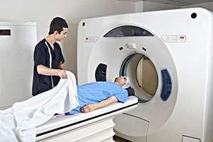
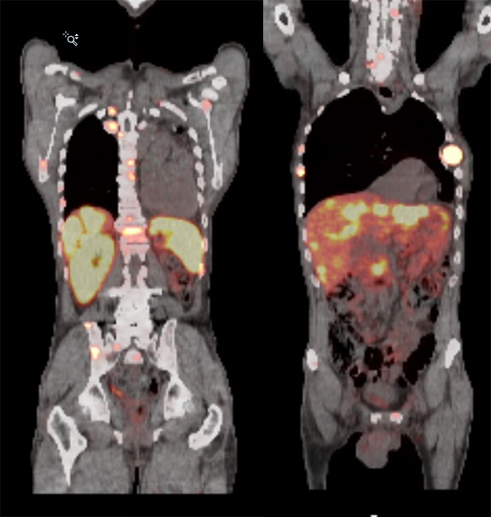
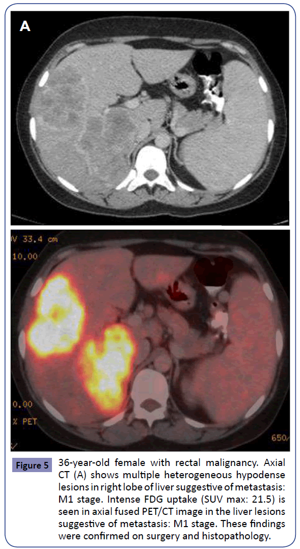
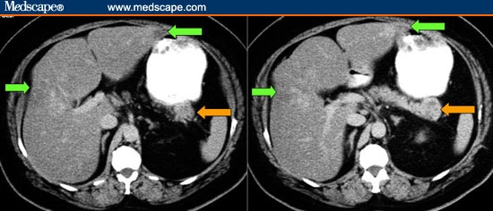
Pet ct scan for liver cancer. One small clinical trial that directly compared psma pet ct with fluciclovine f18 pet ct showed that the psma targeted scan found more metastatic tumors regardless of their location. A ct scan can provide precise information about the size shape and position of tumors in the liver or elsewhere in the abdomen as well as. The pet scan will follow and can take anywhere from 20 to 45 minutes depending on the purpose and scope of the test.
Combining a pet scan with an mri or ct scan can help make the images easier to interpret. The reality is that you cannot rely on a ct scan or ultrasound mri or blood test to tell you if you have cancer. After 13 rounds of chemo my 2nd pet scan yesterday showed nothing in my breast or lymph nodes.
This sugar substance is taken up by cells that use the most energy. Obviously the chemo is working. Sometimes your doctor may refer to a pet ct scan as a pet scan or pet imaging.
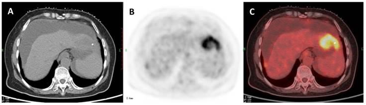
A small amount of a radioactive sugar substance is injected into the patients body. A petct scan is occasionally used for secondary cancer in the liver that has spread from the bowel or from a melanoma. Petct which is a combination of positron emission tomography pet with.
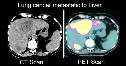
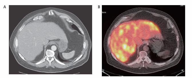
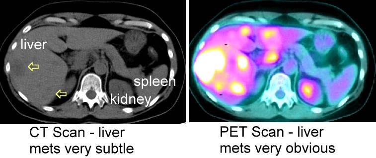
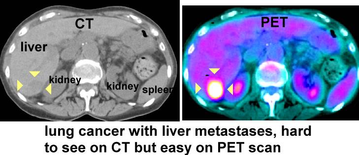
Petct scans provide significantly more information than ct scans and are far more reliable when diagnosing cancer. At left is a ct scan while the center image is from a pet scanner. The ct scan only takes around two minutes.
If you are experiencing bone pain or blood tests reveal elevated calcium levels your radiation oncologist may perform a bone scan to detect whether liver cancer has spread to the bone. 4 months ago my first pet scan showed my breast tumor one lymph node in my armpit and one lymph node in my chest. A pet positron emission tomography scan combined with a ct scan is a specialised imaging test.
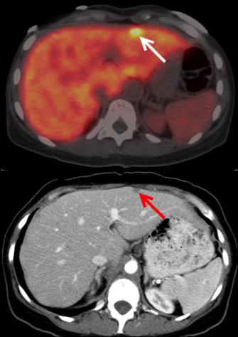
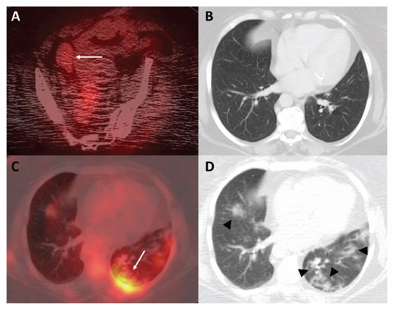
The bright spot in the chest seen best on the pet and ct pet scans is lung cancer. The two scans provide more detailed and accurate information about where some cancers are in the body. Pet ct scan evaluating mestatic liver cancer brendannoyes.
A pet ct scan is a way to create pictures of organs and tissues inside the body. Nci is funding a similar but larger clinical trial. A positron emission tomography pet scan is an imaging test that allows your doctor to check for diseases in your body.
However the doctor was perplexed to see a spot on my liver. The image on the right is a combined ct pet scan. If a pet ct scan is performed the ct scan will be performed first.
Comparing ct and pet ct. My first pet scan in august of 2006 showed high metabolic activity in the hip as was expected and the liver 42 suv.
Immuno Pet Shows Promise For Detecting And Treating Pancreatic Tumors
medicalxpress.com

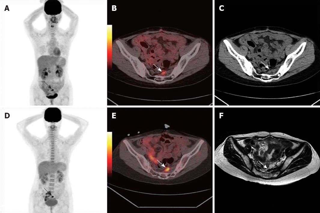
18 F Fdg Pet Ct Images Of Recurrent Ovarian Cancer 10 Mo After Download Scientific Diagram
www.researchgate.net
Tazgknyu9sultm

Registered Axial Images Of A Patient With Liver Metastatic Colorectal Download Scientific Diagram
www.researchgate.net

18 F Fdg Pet Ct Helps Differentiate Autoimmune Pancreatitis From Pancreatic Cancer Bmc Cancer Full Text
bmccancer.biomedcentral.com
Https Encrypted Tbn0 Gstatic Com Images Q Tbn 3aand9gct 5pezfsm2se3gft Izp Vu2hbhg Ujvcvszdkjxpfbrquptks Usqp Cau
encrypted-tbn0.gstatic.com
Clinical Impact Of 18f Fdg Pet Ct On Initial Staging And Therapy Planning For Breast Cancer
www.spandidos-publications.com

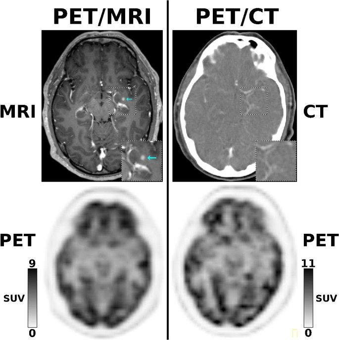
Positron Emission Tomography Magnetic Resonance Imaging Pet Mri An Update And Initial Experience At Hc Fmusp
www.scielo.br
Anatomy Of Liver Mets
www.aboutcancer.com

Secondary Liver Cancer Ct And Pet Scan Stock Image C039 0158 Science Photo Library
www.sciencephoto.com

Secondary Liver Cancer Ct Scan Stock Image C016 6671 Science Photo Library
www.sciencephoto.com
Oncologic Pet By Anatomical Region Radiology Key
radiologykey.com

Basis Of Pet Ct Scan Rgcirc
www.rgcirc.org

Secondary Liver Cancer Ct And Pet Scans Stock Image C016 6770 Science Photo Library
www.sciencephoto.com
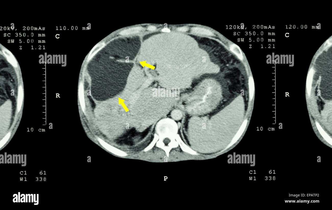
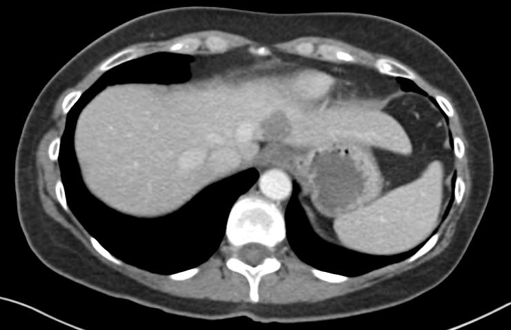
Ct Scan Of Upper Abdomen Show Abnormal Mass At Liver Liver Cancer Stock Photo Alamy
www.alamy.com

Common Causes Of False Positive F18 Fdg Pet Ct Scans In Oncology
www.scielo.br

Upstaging By Fdg Pet In A Patient With Liver Metastasis Of Colon Download Scientific Diagram
www.researchgate.net
Pet Ct In Patients With Liver Lesions Of Different Nature Springerlink
link.springer.com

Added Value Of 18 F Fdg Pet Ct In Patients With Pancreatic Cancer Initial Observation Sciencedirect
www.sciencedirect.com

Pet Ct Open Air Mri Of Cen La
openairmri.com
Pet Imaging 101 Pet Scans
www.itnonline.com
Clinical Value Of 18f Fdg Pet Ct In Assessing Suspicious Relapse After Rectal Cancer Resection
www.wjgnet.com
Psma Pet Ct Accurately Detects Prostate Cancer Spread National Cancer Institute
www.cancer.gov
Pet Ct In Patients With Liver Lesions Of Different Nature Springerlink
link.springer.com
Https Encrypted Tbn0 Gstatic Com Images Q Tbn 3aand9gctu9ril3dcqk Yuo6nxtfwyqzzscolkivc Bq Usqp Cau
A New In Vivo Model To Analyze Hepatic Metastasis Of The Human Colon Cancer Cell Line Hct116 In Nod Shi Scid Il 2rgnull Nog Mice By 18f Fdg Pet Ct
www.spandidos-publications.com
Https Encrypted Tbn0 Gstatic Com Images Q Tbn 3aand9gcrxeuvtqhgb84aysk0i5 D66e9huzaug4tgr Az0o6c838pigk Usqp Cau
encrypted-tbn0.gstatic.com

Liver Cancer 3 Cm Tumour Out 8 Cm Tumour In Ca Care
www.cacare.com

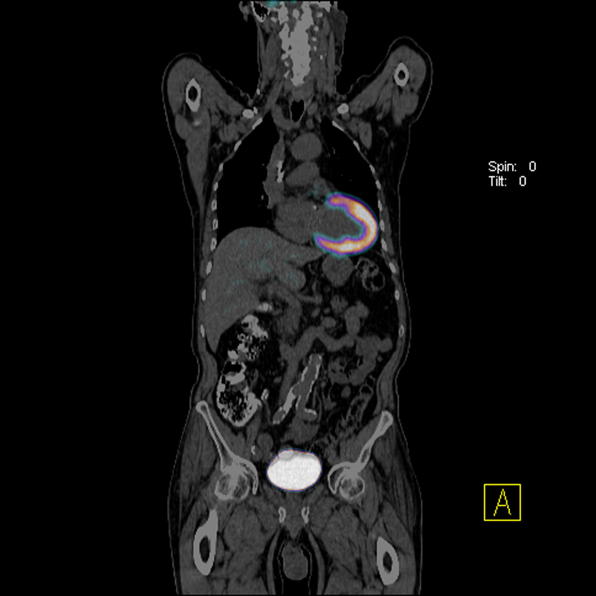
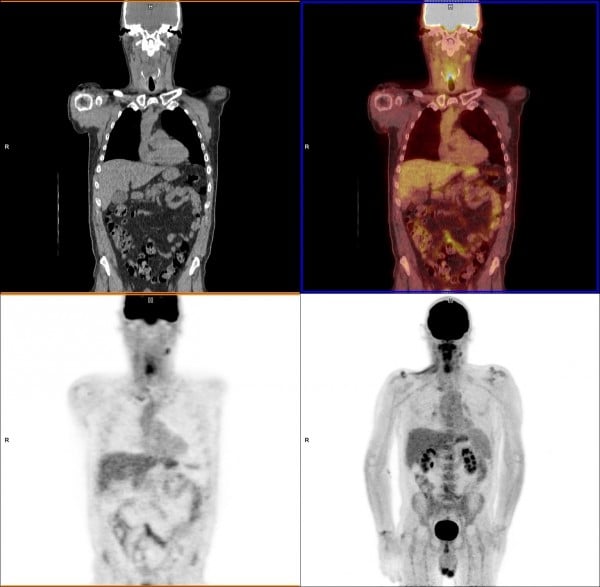
Pet Scan Image Of Whole Body Comparison Axial Coronal Plane In Patient Liver Cancer Recurrence Treatment By Pet Ct Stock Photo Download Image Now Istock
www.istockphoto.com

Pdf Fdg Pet Ct Demonstration Of Metastatic Neuroendocrine Tumor Of Prostate
www.researchgate.net
Pet Mri Versus Pet Ct In Oncology A Prospective Single Center Study Of 330 Examinations Focusing On Implications For Patient Management And Cost Considerations Springerlink
link.springer.com

Fact Sheet Molecular Imaging And Colorectal Cancer Snmmi
www.snmmi.org

Pet Scans In Cancer Cases
www.aboutcancer.com

Case Of The Quarter July 2012
www.nepetimaging.com

Non Small Cell Lung Cancer Fdg Pet Ct Imaging Features A Three Download Scientific Diagram
www.researchgate.net
Pet Ct Breast Cancer
www.numebook.cl

Pet Ct Imaging In Lung Cancer Indications And Findings
www.scielo.br
18f Fdg Pet Ct Can Be Used To Detect Non Functioning Pancreatic Neuroendocrine Tumors
www.spandidos-publications.com

Pet Scans In Cancer Cases
www.aboutcancer.com
How We Read Oncologic Fdg Pet Ct Cancer Imaging Full Text
cancerimagingjournal.biomedcentral.com

Figure 3 From Role Of Pet Ct In The Detection Of Liver Metastases From Colorectal Cancer Semantic Scholar
www.semanticscholar.org
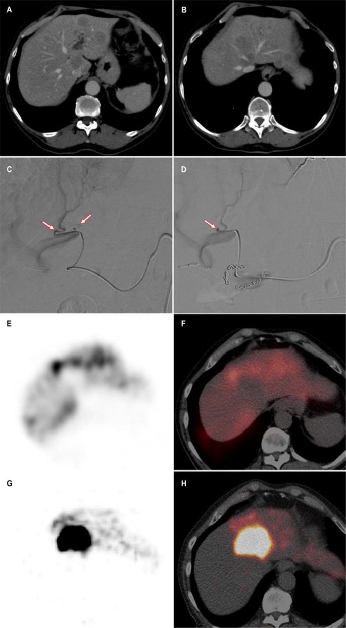
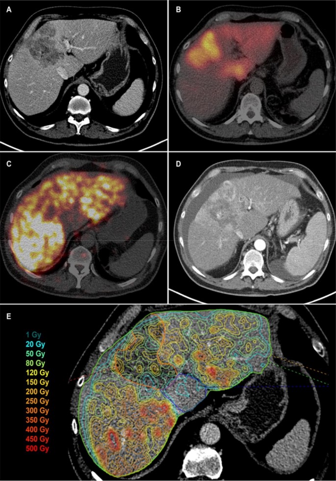
The Value Of Yttrium 90 Pet Ct After Hepatic Radioembolization A Pictorial Essay Springerlink
link.springer.com

Staging Pet Mri Scan Of A 56 Year Old Woman With Known Ovarian Cancer Download Scientific Diagram
www.researchgate.net
Staging Performance Of Whole Body Dwi Pet Ct And Pet Mri In Invasive Ductal Carcinoma Of The Breast
www.spandidos-publications.com

Positron Emission Tomography Magnetic Resonance Imaging Pet Mri An Update And Initial Experience At Hc Fmusp
www.scielo.br
The Value Of Yttrium 90 Pet Ct After Hepatic Radioembolization A Pictorial Essay Springerlink
link.springer.com
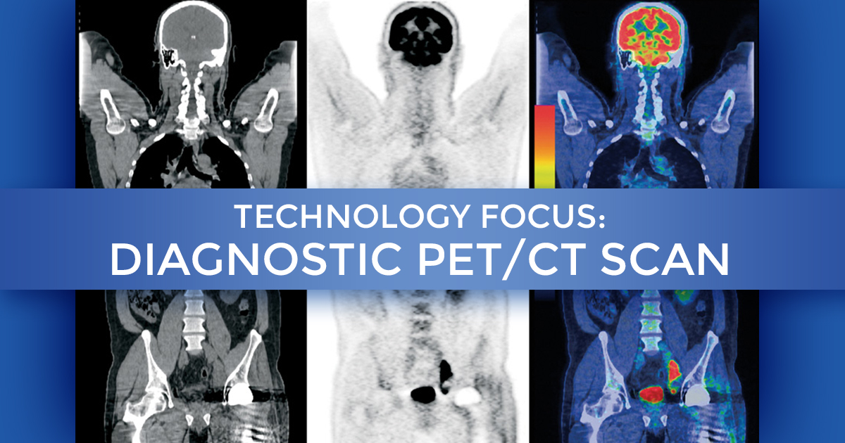
Technology Focus Diagnostic Pet Ct Scan At Summit Cancer Centers
www.summitcancercenters.com

Breast Cancer Cancer Probe
sites.google.com

Pet Ct
www.med-ed.virginia.edu

Typical Example Of Colorectal Cancer Patient With Nonresectable Liver Download Scientific Diagram
www.researchgate.net

Pet Ct Scan Liver Cancer Axial Stock Illustration 1282563898
www.shutterstock.com
Plos One Hepatic Lesions Detected After Mastectomy In Breast Cancer Patients With Hepatitis Background May Need To Undergo Liver Biopsy To Rule Out Second Primary Hepatocellular Carcinoma
journals.plos.org

A Initial Positron Emission Tomography Computed Tomography Pet Ct Download Scientific Diagram
www.researchgate.net
Https Encrypted Tbn0 Gstatic Com Images Q Tbn 3aand9gcqtnbhpuprprduowp3lroneprmpr6wctflo35qq5vtjl5lt4j40 Usqp Cau
encrypted-tbn0.gstatic.com

Breast Cancer Staging Physiology Trumps Anatomy
www.sbi-online.org
Comparison Of Contrast Enhanced Mdct And Integrated Fdg Pet Ct Staging Of Colorectal Cancer Insight Medical Publishing
colorectal-cancer.imedpub.com

18f Fdg Pet Ct Imaging Of The Pancreas Spectrum Of Diseases Insight Medical Publishing
pancreas.imedpub.com

Staging Psma Pet Ct Scan Showing The Supposed Rapid Prostate Cancer Download Scientific Diagram
www.researchgate.net
Https Encrypted Tbn0 Gstatic Com Images Q Tbn 3aand9gcqejzagkr3f Dth Xesrswsh3z4c58sd3vydefffgponakcwcpk Usqp Cau
encrypted-tbn0.gstatic.com

Advantages Of Pet Ct In Staging Lung Cancer Coronal Slice Of A Pet Ct Download Scientific Diagram
www.researchgate.net

Pet Ct Scan In The Staging Of Left Breast Cancer Detection Performed 1 Download Scientific Diagram
www.researchgate.net

Is Radiation From A Ct Or Pet Scan Dangerous Cancer Ut Southwestern Medical Center
utswmed.org

Image Registration
www.ucl.ac.uk
Incidental Covid 19 On Pet Ct Imaging Cmaj
www.cmaj.ca

Pet Scans In Cancer Cases
www.aboutcancer.com
The New Powers Of Pet Ct Scan Technology San Cristobal Cancer Institute
sancristobalcancer.com

Pet Imaging In Hematology Ask Hematologist Understand Hematology
askhematologist.com
Pet Imaging In Hematology Ask Hematologist Understand Hematology
askhematologist.com
Differentiation Between Malignant And Benign Solitary Lesions In The Liver With 18fdg Pet Ct Accuracy Of Age Related Diagnostic Standard
www.jcancer.org

Positron Emission Tomography Computed Tomography In The Management Of Lung Cancer An Update Sharma P Singh H Basu S Kumar R South Asian J Cancer
journal.sajc.org

Neuroendocrine Cancer If You Can See It You Can Detect It Ronny Allan Living With Neuroendocrine Cancer
ronnyallan.net

Secondary Liver Cancer Ct And Pet Scans Stock Image C001 7975 Science Photo Library
www.sciencephoto.com

2 8 F Fdg Pet Ct Scan Showing Three Liver Metastases 12 Months After Download Scientific Diagram
www.researchgate.net

Lung Cancer Snmmi
www.snmmi.org

18f Fdg Pet Ct Imaging Of Colorectal Cancer A Pictorial Review Postgraduate Medical Journal
pmj.bmj.com

Pet Ct Scan Of Patient With Pulmonary Metastases Of Rectal Cancer Top Download Scientific Diagram
www.researchgate.net

Secondary Liver Cancer Ct And Pet Scan Stock Image C016 6768 Science Photo Library
www.sciencephoto.com
Pet Scans In Cancer Cases
www.aboutcancer.com

The Role Of Pet Scans In The Diagnosis And Treatment Of Cancer
news.cancerconnect.com
Pet Ct And Pet Mri In Ophthalmic Oncology Review
www.spandidos-publications.com

Patient With Breast Cancer Both Pet Ct A B As Well As Pet Mri C D Download Scientific Diagram
www.researchgate.net
Assessment Of Liver Lesions
www.medscape.org
Pet Scans In Cancer Cases
www.aboutcancer.com

Sirtex Diagnosis Of Liver Tumors
www.sirtex.com

Secondary Liver Cancer Ct And Pet Scan Stock Image C016 6771 Science Photo Library
www.sciencephoto.com

Liver Cancer Ct Scan Stock Image C010 4902 Science Photo Library
www.sciencephoto.com
Secondary Liver Tumors Intechopen
www.intechopen.com
Plos One Hepatic Lesions Detected After Mastectomy In Breast Cancer Patients With Hepatitis Background May Need To Undergo Liver Biopsy To Rule Out Second Primary Hepatocellular Carcinoma
journals.plos.org

Pet Scans In Dallas Tx Positron Emission Tomography
swdcmi.com
Pet Ct In Patients With Liver Lesions Of Different Nature Springerlink
link.springer.com

Application Of Different Imaging Methods In The Early Diagnosis Of Primary Hepatic Carcinoma
www.hindawi.com

Liver Metastases Pet Ct Scan Stock Image C017 4432 Science Photo Library
www.sciencephoto.com

Pet Ct Imaging In Lung Cancer Indications And Findings
www.scielo.br
The Role Of 18f Fdg Pet Ct Integrated Imaging In Distinguishing Malignant From Benign Pleural Effusion
journals.plos.org

Colorectal Cancer Patterns Of Locoregional Recurrence And Distant Metastases As Demonstrated By Fdg Pet Ct Purandare Nc Dua Sg Arora A Shah S Rangarajan V Indian J Radiol Imaging
www.ijri.org
Figure 1 From Role Of Pet Ct In The Detection Of Liver Metastases From Colorectal Cancer Semantic Scholar
www.semanticscholar.org

Liver Cancer 3 Cm Tumour Out 8 Cm Tumour In Ca Care
www.cacare.com

1 Verdict Hospital
www.hospitalmanagement.net
