Pet Ct Scan Lung Cancer Images
Integrated Pet Ct In The Staging Of Nonsmall Cell Lung Cancer Technical Aspects And Clinical Integration European Respiratory Society
erj.ersjournals.com

Figure 1 From Pet In Lung Cancer Semantic Scholar
www.semanticscholar.org

Pet Ct Scan Axial Fusion Images Showing Fdg Avid Right Upper Lobe Lung Download Scientific Diagram
www.researchgate.net
Researchers Recommend Additional Pet Ct Scans In Lung Cancer Follow Up
lungdiseasenews.com

Staging Of Non Small Cell Lung Cancer With Integrated Positron Emission Tomography And Computed Tomography Nejm
www.nejm.org

Role Of 18f Fdg Pet In Assessment Of Response In Non Small Cell Lung Cancer
jnm.snmjournals.org
The bronchi are the airways that carry air to the lungs and mouth.

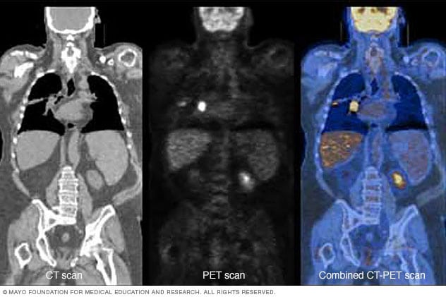
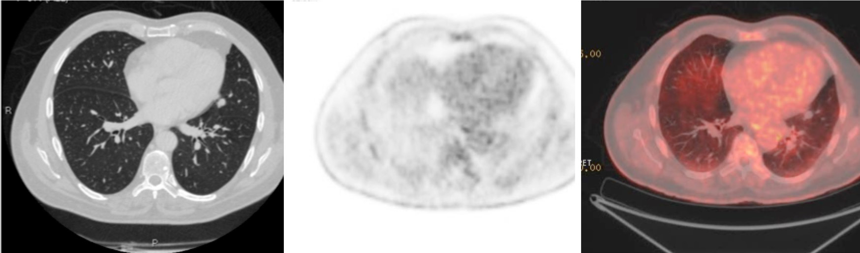
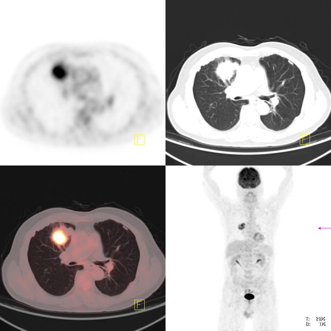
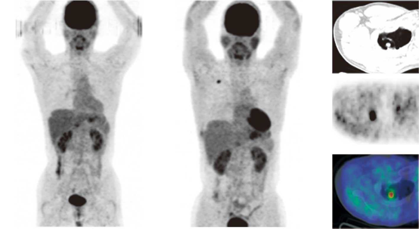
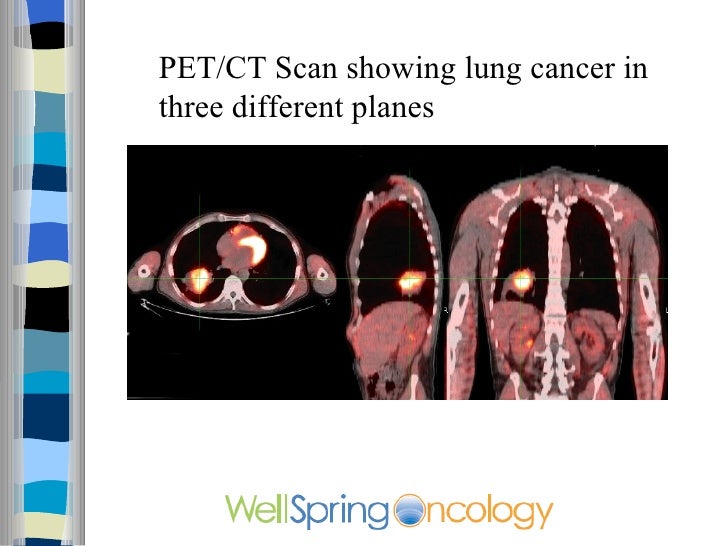
Pet ct scan lung cancer images. It is sometimes called computerized tomography or computerized axial tomography cat. Fluoro deoxyglucose positron emission tomography pet imaging has a diagnostic and prognostic value in the initial staging restaging and surveillance of nonsmall cell lung cancer nsclc. At left is a ct scan while the center image is from a pet scanner.
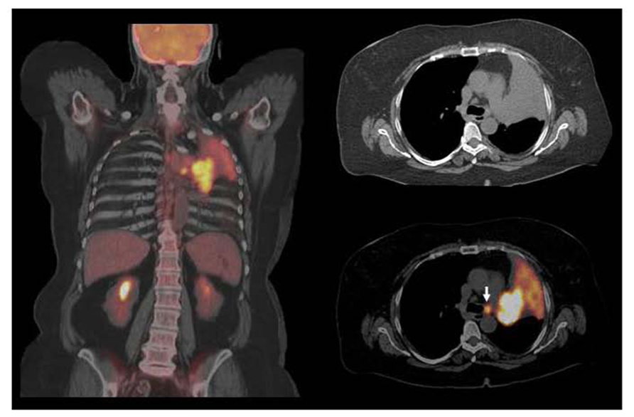
A lung pet scan is typically combined with a lung ct scan to detect conditions like lung cancer. The image on the right is a combined ct pet scan. Each picture created during a ct procedure.
It can give more information about any abnormalities nodules or lesions small abnormal areas in the lungs that were. A ct scan takes a cross sectional and a more detailed image of the lung. How the test is performed.
Coloured computed tomography ct scan of a section through the chest of a 76 year old male patient with a malignant cancerous tumour dark right of the bronchus. You can usually drink water during this time. Unlike conventional x rays ct scans provide exceptionally detailed images of the bones organs and tissuesx rays are taken from many angles and combined to create a cross sectional image.
The term tomography comes from the greek words tomos a cut a slice or a section and graphein to write or record. Unlike magnetic resonance imaging mri and computed tomography ct scans which reveal the structure of the lungs a pet scan shows how well the lungs and their tissues are working. Combining a pet scan with an mri or ct scan can help make the images easier to interpret.
Like ct scans they can produce detailed images of the tissue in the chest cavity. When used in conjunction with conventional radiologic imaging pet imaging has been shown to result in significant changes in clinical management of nsclc. They are most often used to see if lung cancer has spread beyond its initial site.
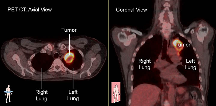
Pet scans which use fluorodeoxyglucose fdg injected into the body to illuminate cancer cells. Computed tomography is an imaging procedure that uses special x ray equipment to create detailed pictures or scans of areas inside the body. It uses a radioactive substance called a tracer to look for disease in the lungs such as lung cancer.
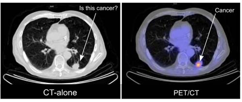
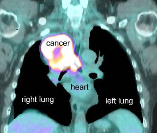
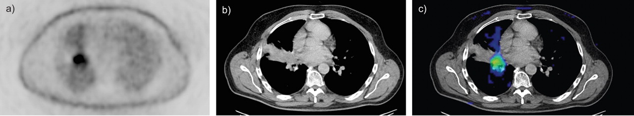
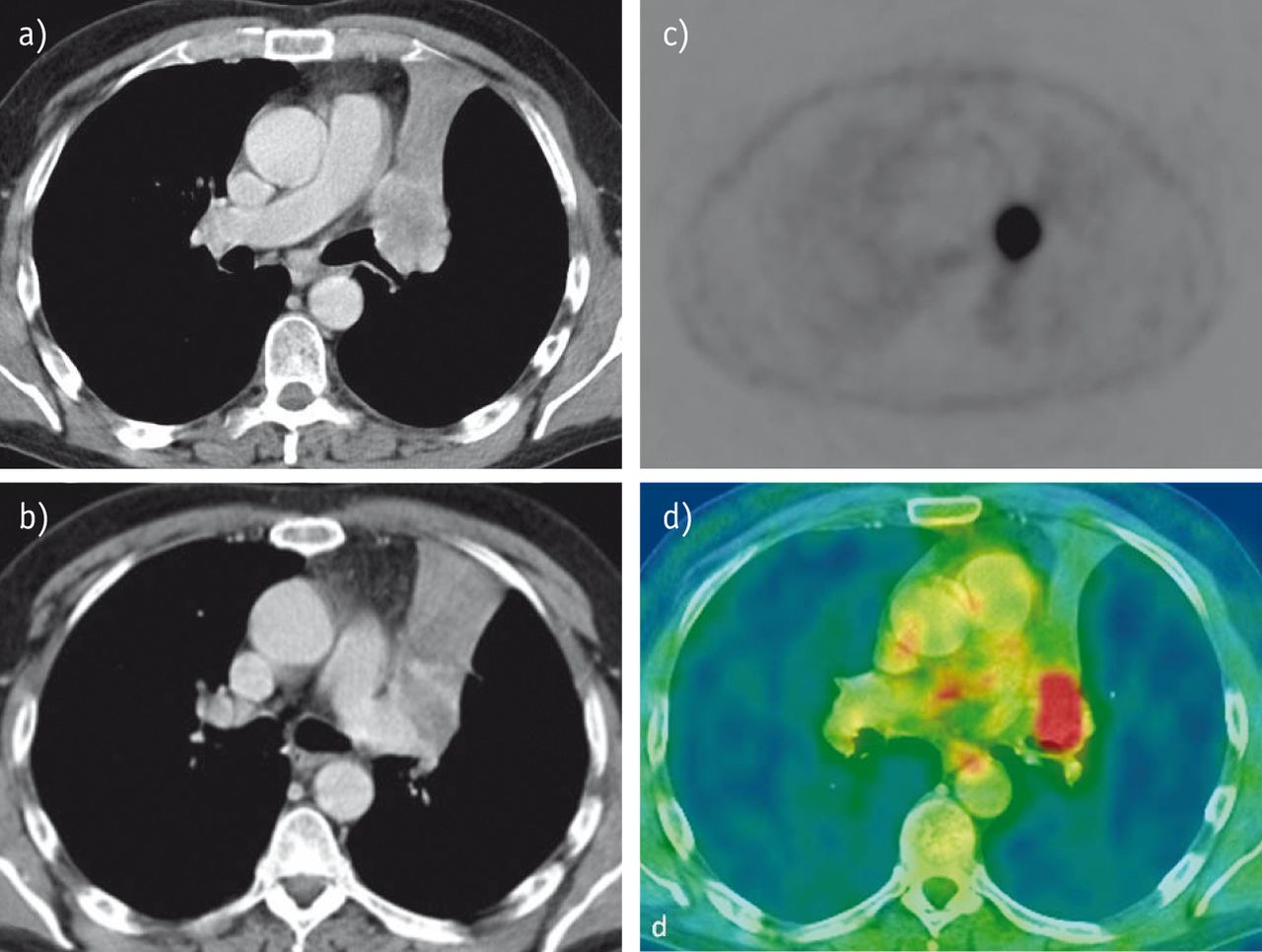
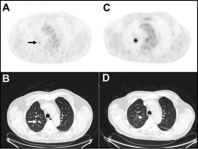
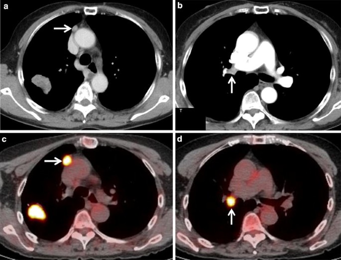
Preparing for your pet ct scan for most pet ct scans you need to stop eating about 4 to 6 hours beforehand. The bright spot in the chest seen best on the pet and ct pet scans is lung cancer. Its also useful in determining if cancer has spread beyond the initial site.
Application Of Pet Ct In Lung Cancer Pet Ct Imaging Of Berkeley
petctberkeley.com
A Case Of Primary Lung Cancer Lesion Demonstrated By F 18 Fdg Positron Emission Tomography Computed Tomography Pet Ct One Year After The Detection Of Metastatic Brain Tumor
www.spandidos-publications.com

18f Fdg Pet Ct Imaging Of The Pancreas Spectrum Of Diseases Insight Medical Publishing
pancreas.imedpub.com
Https Encrypted Tbn0 Gstatic Com Images Q Tbn 3aand9gcqscm8qlx9qwapcwilybirptagvuhn1elwhse0vz1jsa Jtsfgq Usqp Cau
encrypted-tbn0.gstatic.com
Pet Ct Scans Showed The Mass With Increased 18 Fdg Uptake In Left Main Download Scientific Diagram
www.researchgate.net

Pet Ct Scan Image Of Whole Body Comparison Axial Plane In Ct Scan And Pet Ct For Detect Cancer Recurrence In Patient Lung Cancer Disease Stock Photo Download Image Now Istock
www.istockphoto.com
Differentiation Of Central Lung Cancer From Atelectasis Comparison Of Diffusion Weighted Mri With Pet Ct
journals.plos.org
Pet Plus Ct Mayo Clinic
www.mayoclinic.org

View Image
www.ijem.in

Positron Emission Tomography Computed Tomography In The Management Of Lung Cancer An Update Sharma P Singh H Basu S Kumar R South Asian J Cancer
journal.sajc.org

Pet Ct Open Air Mri Of Cen La
openairmri.com
Xrays And Ct Scans Of Lung Cancer
www.aboutcancer.com
Psma Pet Ct Accurately Detects Prostate Cancer Spread National Cancer Institute
www.cancer.gov
Imaging Of Solitary Pulmonary Nodule A Clinical Review Sim Quantitative Imaging In Medicine And Surgery
qims.amegroups.com
Https Encrypted Tbn0 Gstatic Com Images Q Tbn 3aand9gcrfi1fyvtwbrhsvbvdyoi302r97l3jii2qmwzp7cjqxzoxtdvmq Usqp Cau
encrypted-tbn0.gstatic.com
Pet Ct Tracer Identifies Vulnerable Lesions In Non Small Cell Lung Cancer Patients Imaging Technology News
www.itnonline.com

Science Source Pet Ct Scan Of Lung Cancer
www.sciencesource.com
Imaging Tests Lungevity Foundation
lungevity.org

Ground Glass Opacity Lung Nodules In The Era Of Lung Cancer Ct Screening Radiology Pathology And Clinical Management
www.cancernetwork.com

New Diagnostic Test For The Early Detection Of Lung Cancer Dr Robert A Nagourney Rational Therapeutics Blog
robertanagourney.wordpress.com

18f Fdg Pet Ct Imaging Of The Pancreas Spectrum Of Diseases Insight Medical Publishing
pancreas.imedpub.com

Pet Ct Scan Where Can You Get Pet Ct Scan Done At Affordable Price
blog.chuzdr.com
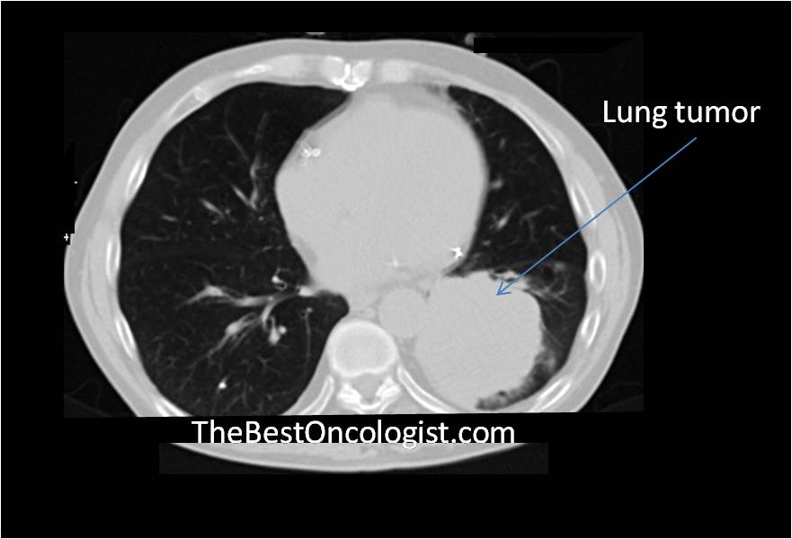
How Lung Cancer Is Diagnosed The Best Oncologist Tm
www.thebestoncologist.com
Pet Ct Principles And Applications In Lung Cancer Management Intechopen
www.intechopen.com
The New Powers Of Pet Ct Scan Technology San Cristobal Cancer Institute
sancristobalcancer.com

Ct And Pet Lung Cancer
ykhoa.org

Pulmonary Aspergilloma A Rare Differential Diagnosis To Lung Cancer After Positive Fdg Pet Scan Sciencedirect
www.sciencedirect.com
Clinical Impact Of 18f Fdg Pet Ct On Initial Staging And Therapy Planning For Breast Cancer
www.spandidos-publications.com
Advanced Radiology Consultants Pet Ct
www.adrad.com

Pet Ct Scan Of Lung Cancer Yoga Mat For Sale By Living Art Enterprises Llc
fineartamerica.com
Present And Future Roles Of Fdg Pet Ct Imaging In The Management Of Lung Cancer Springerlink
link.springer.com

Updated Lung Cancer Screening Guidelines Released At Chest Pulmonology Advisor
www.pulmonologyadvisor.com

Pet Scan Images Lung Cancer Cancer News Update
popcultureworldnews.com

Non Small Cell Lung Cancer Fdg Pet Ct Imaging Features A Three Download Scientific Diagram
www.researchgate.net

Small Cell Lung Cancer Chest Ct Scan A And Corresponding Pet Ct Download Scientific Diagram
www.researchgate.net
Https Encrypted Tbn0 Gstatic Com Images Q Tbn 3aand9gcq6xjxerjxit4qxz Awgdqgljwdo9 5aocltsa6ftfkc5tdeu2q Usqp Cau
encrypted-tbn0.gstatic.com

Non Small Cell Lung Cancer Fdg Pet Ct Imaging Features A Three Download Scientific Diagram
www.researchgate.net
A Case Of Primary Lung Cancer Lesion Demonstrated By F 18 Fdg Positron Emission Tomography Computed Tomography Pet Ct One Year After The Detection Of Metastatic Brain Tumor
www.spandidos-publications.com

Research Points To Value For Fourth Follow Up Pet Ct Scans For Lung Cancer Patients
axisimagingnews.com

Analysis Of Murine Lung Tumors By Micro Pet Ct Imaging
bio-protocol.org

Positron Emission Tomography Computed Tomography In The Management Of Lung Cancer An Update
journal.sajc.org

Image Guided Radiation Therapy For Non Small Cell Lung Cancer Sciencedirect
www.sciencedirect.com
False Positive Finding From Malignancy Like Lesions On Fdg Pet Ct Case Report Of Tuberculosis Patients Bmc Medical Imaging Full Text
bmcmedimaging.biomedcentral.com

Pet Ct Imaging In Lung Cancer Indications And Findings
www.scielo.br

Xrays And Ct Scans Of Lung Cancer
www.aboutcancer.com

1 Verdict Hospital
www.hospitalmanagement.net

Lung Cancer Ct And Pet Scan Stock Image C016 6772 Science Photo Library
www.sciencephoto.com
Integrated Pet Ct In The Staging Of Nonsmall Cell Lung Cancer Technical Aspects And Clinical Integration European Respiratory Society
erj.ersjournals.com

Non Small Cell Lung Cancer Ct Pet Scan Stock Image C007 1648 Science Photo Library
www.sciencephoto.com
Imaging Techniques In Lung Cancer European Respiratory Society
breathe.ersjournals.com

Pet Ct Imaging In Lung Cancer Indications And Findings
www.scielo.br
Https Encrypted Tbn0 Gstatic Com Images Q Tbn 3aand9gctbt6avfwdwc7 Kcxtlsd5blbqzxqeqszgp2olleszshofk78y0 Usqp Cau
encrypted-tbn0.gstatic.com

Pet Ct
www.med-ed.virginia.edu

Positron Emission Tomography Pet Snmmi
www.snmmi.org

Pet Scans In Cancer Cases
www.aboutcancer.com

Imaging Of Lung Cancer Implications On Staging And Management Purandare Nc Rangarajan V Indian J Radiol Imaging
www.ijri.org

Pet Ct Imaging In Lung Cancer Indications And Findings
www.scielo.br
Diagnostics Free Full Text Early Response Assessment To Targeted Therapy Using 3 Deoxy 3 18 F Fluorothymidine 18f Flt Pet Ct In Lung Cancer Html
www.mdpi.com

Lung Cancer Snmmi
www.snmmi.org

18f Fdg Pet Ct And Lung Cancer Value Of Fourth And Subsequent Posttherapy Follow Up Scans For Patient Management
jnm.snmjournals.org
Understanding Your Fdg Pet Scan
www.docpanel.com

Pet Ct Imaging For Target Volume Delineation In Curative Intent Radiotherapy Of Non Small Cell Lung Cancer Iaea Consensus Report 2014 Radiotherapy And Oncology
www.thegreenjournal.com
For Early Stage Lung Cancer A New Biomarker And Treatment Target National Cancer Institute
www.cancer.gov

Fdg Pet Ct As Theranostic Imaging In Diagnosis Of Non Small Cell Lung Cancer
www.bioscience.org

Pet Ct Scan Image Of Whole Body Comparison Axial Plane In Ct Scan And Pet Ct On The Screen For Detect Cancer Recurrence In Patient Lung Cancer Disease Stock Photo Download
www.istockphoto.com
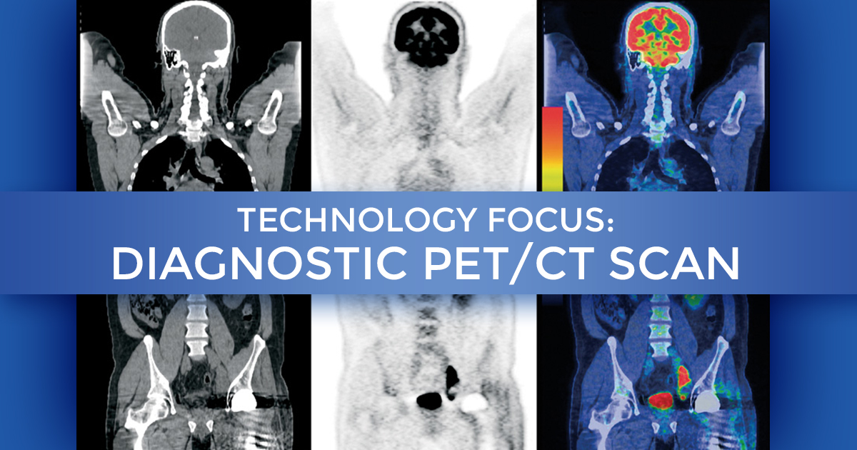
Technology Focus Diagnostic Pet Ct Scan At Summit Cancer Centers
www.summitcancercenters.com

Pet Ct
www.med-ed.virginia.edu

Health Victoria Pet Ct Scanning Leads To Guided Lung Cancer Therapy May 2013
www.health.vic.gov.au
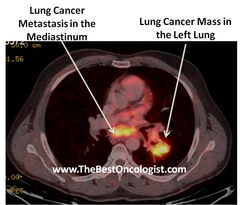
How Lung Cancer Is Diagnosed The Best Oncologist Tm
www.thebestoncologist.com

Lung Cancer Ct And Pet Scans Stock Image C010 3488 Science Photo Library
www.sciencephoto.com

Inclusion Of Brain In Fdg Pet Ct Scanning Techniques In Cancer Patients Does It Obviate The Need For Dedicated Brain Imaging Semantic Scholar
www.semanticscholar.org
Differentiation Of Central Lung Cancer From Atelectasis Comparison Of Diffusion Weighted Mri With Pet Ct
journals.plos.org

Gracecast 015 Cancer 101 David Djang Interview On Pet Scans Youtube
www.youtube.com
Stock Image Ct Scan Pet Scan And Petct Fusion Image Of The Body Showing Osseous Bony Cancer Of The D6 Dorsal And L5 Lumbar Vertebrae Metastases Of Lung Cancer Frontal Sections Petct
www.medicalimages.com
Pet Ct Principles And Applications In Lung Cancer Management Intechopen
www.intechopen.com

Role Of Positron Emission Tomography Computed Tomography In Carcinoma Lung Evaluation Padma S Sundaram P S George S J Can Res Ther
www.cancerjournal.net
Researchers Recommend Additional Pet Ct Scans In Lung Cancer Follow Up
lungdiseasenews.com

Lung Cancer Clinical Advisor
www.clinicaladvisor.com

Lung Cancer Pet And 3d Ct Scan Stock Image C026 7601 Science Photo Library
www.sciencephoto.com
Present And Future Roles Of Fdg Pet Ct Imaging In The Management Of Lung Cancer Springerlink
link.springer.com

Integrated Pet Ct In The Staging Of Nonsmall Cell Lung Cancer Technical Aspects And Clinical Integration European Respiratory Society
erj.ersjournals.com
Lung Cancer
es.slideshare.net

Pet Ct Scan Vs Ct Scan For Cancer Diagnosis
www.ctoam.com

Pulmonary Cryptococcosis Mimicking Solitary Lung Cancer In An Immunocompetent Patient Thorax
thorax.bmj.com
The Role Of 18f Fdg Pet Ct Integrated Imaging In Distinguishing Malignant From Benign Pleural Effusion
journals.plos.org

A Representative Case Of 18 F Fdg Pet Ct For Lymph Node Staging In Lung Download Scientific Diagram
www.researchgate.net

Case Of The Quarter July 2011
www.nepetimaging.com
Plos One 68ga Dotatate Pet Ct Imaging Of Indeterminate Pulmonary Nodules And Lung Cancer
journals.plos.org

What Is Pet Ct Scan Get A Detailed Overview Checkout The Real Cost Of Pet Ct Scan By Bookmyscan Bangalore Issuu
issuu.com

Fact Sheet Molecular Imaging And Lung Cancer Snmmi
www.snmmi.org
The Role Of Pet Ct In Detecting Bone Metastases In Patients With Breast Cancer Site Title
hwilczega.wordpress.com

Fdg Pet Ct For Solitary Pulmonary Nodule And Lung Cancer Literature Review Sciencedirect
www.sciencedirect.com

Xrays And Ct Scans Of Lung Cancer
www.aboutcancer.com

Screening For Lung Cancer Takes A Lot Of Effort To Find A Small Number Of Cancers Shots Health News Npr
www.npr.org
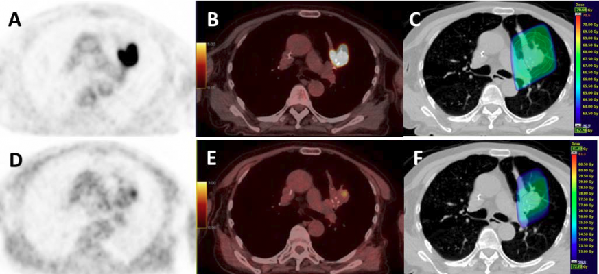
Frontiers Current Concepts In F18 Fdg Pet Ct Based Radiation Therapy Planning For Lung Cancer Oncology
www.frontiersin.org

Ct Scan Computed Tomography Scan Cat Scan Pet Scan Lung Cancer
computedtomographyscan.blogspot.com
Additional Pet Ct Scans Valuable In Follow Up Of Lung Cancer Patients Imaging Technology News
www.itnonline.com

Pet Ct Imaging In Lung Cancer Indications And Findings
www.scielo.br
